급성호흡곤란증후군으로 나타난 NK-T 세포 림프종
NK-T cell lymphoma manifesting as acute respiratory distress syndrome
Article information
Abstract
양측성 폐침윤을 보인 49세 여자에서 항생제 복합투여에도 상태가 더욱 악화되고 급성호흡곤란증후군으로 진행하였다. 폐병변의 원인을 밝히기 위하여 기관지 폐포 세척 및 폐생검을 시행하였고, NK-T 세포 림프종을 진단하여 보고하는 바이다.
Trans Abstract
Primary pulmonary lymphoma is a rare disease, and non-B cell lymphomas (T-cell and natural killer cell lymphomas) involving the lung parenchyma are uncommonly reported. The most common radiological feature of pulmonary parenchymal lymphoma is a single mass or nodule. A 49-year-old woman with dyspnea was referred with suspicion of severe pneumonia. A chest radiograph showed diffuse nodular infiltration in both lungs. Acute respiratory failure was severe and rapidly progressive, so she was managed with a mechanical ventilator under the impression of acute respiratory distress syndrome (ARDS). A bronchoalveolar lavage and lung biopsy by video-assisted thoracic surgery revealed NK-T cell lymphoma. We report a case of extranodal NK-T cell lymphoma presenting as ARDS. (Korean J Med 79:697-700, 2010)
서 론
NK-T 세포 림프종은 미국, 유럽에서 매우 드문 질환이나 아시아에서는 좀더 흔하게 나타나고 있다1). 코, 코인두, 입인두, 입천장, 상기도, 위장관, 피부 등과 같은 림프절 외 부위에 잘 생기는 것으로 보고되고 폐실질 침범은 드물게 나타나고 있다1). NK-T 세포 림프종은 공격적인 임상경과를 보여 진단이 늦어질 경우에는 치명적인 결과를 초래할 수 있다. 저자들은 49세 여자에게서 양측성 폐침윤으로 나타나 급성호흡곤란증후군을 보인 NK-T 세포 림프종을 진단하여 보고하는 바이다.
증 례
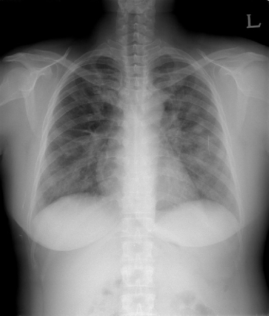
49세 여자가 호흡곤란으로 내원하였다. 내원 10일 전부터 근육통, 열감이 있었고, 7일 전부터 기침, 가래 및 호흡곤란이 있어 개인병원에서 감기약을 복용하였으나 증상이 지속되었다. 당시 시행한 혈액검사 및 흉부 단순촬영에서 중증 폐렴 및 범혈구감소증 소견을 보여 본원으로 전원되었다. 과거병력에서 특이사항은 없었으며 가정주부이고, 흡연과 음주는 하지 않았다. 신체진찰에서 혈압 100/60 mmHg, 맥박 100회/분, 호흡수 24회/분, 체온 37.4℃였고, 의식은 명료하였다. 입술은 청색증은 없었으나, 양측 폐야에서 거친 호흡음 및 마찰음이 들렸다. 대기중 동맥혈가스분석에서 pH 7.49, PaCO2 29.9 mmHg, PaO2 61.8 mmHg, HCO3¯ 22 mmol/L, 산소포화도 93%였다. 말초 혈액검사에서 백혈구 2,400/mm3 (호중구 85.2%, 림프구 9.2%, 단핵구 2.8%), 혈색소 10.9 g/dL, 혈소판 92,000/mm3이었고, AST 136 IU/L, ALT 107 IU/L, 알부민 2.6 g/dL, 프로트롬빈시간 84초(INR 1.13), aPTT 52.1 sec이었다. 흉부단순촬영에서 양쪽 폐 전체에 경계가 명확하지 않은 침윤성 결절들이 있었다(그림 1). 흉부 전산화단층촬영에서 양쪽 폐전체에 소엽 사이막 비후를 동반한 다발성 결절들이 있었고, 기관지옆 림프절, 대동맥활밑 림프절 종대가 있어 세균성 폐렴 및 진균성 폐렴, 폐결핵을 의심하였다(그림 2). 항생제로 cefotaxime 6 g/일, azithromycin 500 mg/일을 사용하였고, fluconazole 400 mg/일을 병용 투여하였다.

Chest CT scan showed multiple ill-defined nodules with interlobular septal thickening in both lung fields.
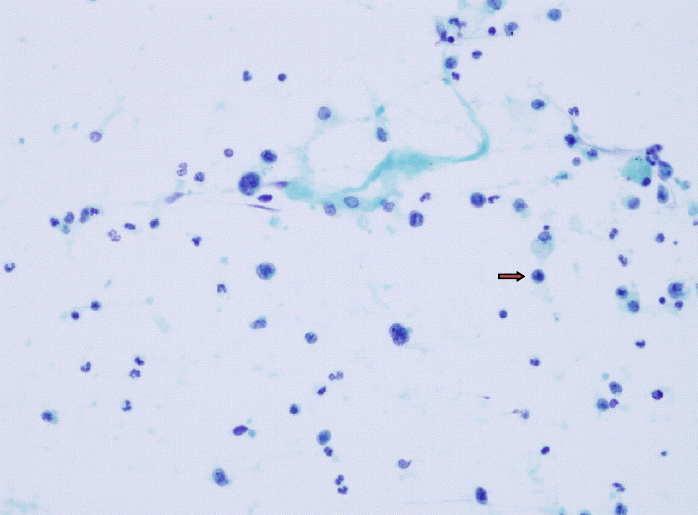
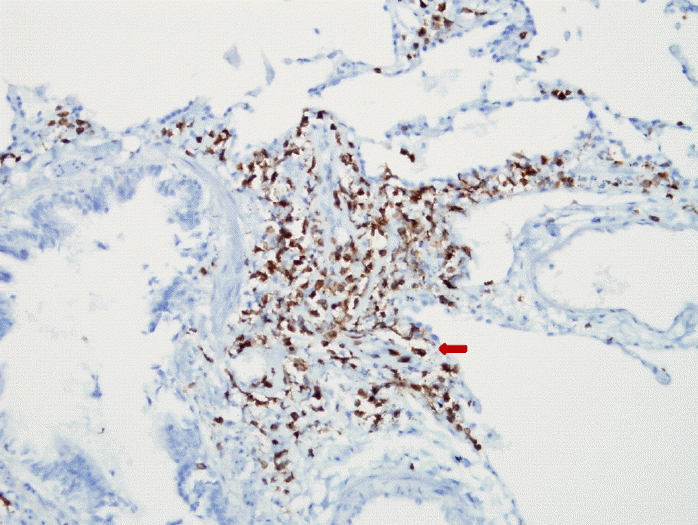
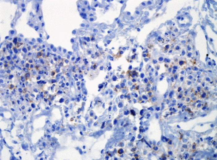
입원 3일 후에도 체온이 38.5℃ 이상 지속되어 항생제 반응이 없는 것으로 판단하여 cefepime 2 g/일, ciprofloxacin 400 mg/일으로 변경하였다. 입원 5병일째에 산소포화도 85%로 감소하며 급성호흡곤란증후군으로 진행하여 기계호흡을 시작하였고, 원인 균주를 확인하기 위하여 기관지 폐포세척을 시행하였다. 폐포 세척액 검사에서 적혈구 3,750/mm3, 백혈구 280/mm3 (호중구 40%, 림프구 16%, 단핵구 27%)였으며, 의미있는 세균, 항산균 및 진균은 검출되지 않았다. 8병일째에 흉부단순촬영에서 진행양상을 보여 methylprednisolone 62.5 mg씩 6시간마다 7일간 투여하였다. 부신피질호르몬 투여 후 5일째 기관탈관을 하고 호흡곤란이 호전되었다. 11병일째에 기관지 폐포 세척액 세포 도말 검사에서 악성 림프 세포가 확인되었다(그림 3). 17병일째에 시행한 흉강경하 폐 및 종격동 림프절 조직검사에서 폐포 종격에 침윤한 악성 림프 세포가 보였고, 면역화학 염색에서 CD3, CD56 양성으로 NK-T 세포 림프종을 진단하였다(그림 4, 5). 이후 항암화학요법을 시행하려 하였으나 21병일째에 장기부전이 급속히 진행되었고, 이틀 후 사망하였다.

Bronchoalveolar lavage cytology showed singly scattered medium to large sized malignant lymphoid cells (arrow, ×400).

Immunochemistry showing that the malignant cells stained CD3. Bronchoalveolar walls to alveolar septae are massively invaded by small to medium sized malignant cells (×200).
고 찰
원발 폐 림프종은 드물게 나타나는 질환이며 원발 폐종양의 0.5%, 림프절외 림프종의 3%를 차지한다. 그 중 B세포 기원 림프종이나 MALT (mucosa associated lymphoid tissue)가 대부분을 차지하고 있으며 Non-B 세포 림프종(T 세포, natural killer 세포 림프종)은 매우 드물게 보고되고 있다2). 대부분 노년층에서 나타났으며 남녀 비율은 대략 1:2 비율을 보였다. 대부분의 증례에서 기침과 호흡곤란이 주 증상으로 나타났으며, 흉부단순촬영에서 양쪽 폐야에 미만성 결절을 보였다. 그 외에 종물처럼 보이는 음영, cryptogenic organizing pneumonia와 같은 병변, hilar 림프절종대, 흉막삼출 등의 소견을 보였다3). 이처럼 임상양상과 방사선학적 소견만으로 정확한 진단이 어려울 때 병리학적 진단이 치료 방향 결정에 도움을 줄 수 있으며 이를 위하여 기관지 폐포 세척술 또는 개흉폐생검 등의 침습적 방법을 시도해야 할 필요가 있다4). 국내에서 원발 폐 NK-T세포 림프종은 정 등5)의 보고가 있지만, 급성호흡곤란으로 진행하여 기관지폐포세척액검사로 악성 림프종을 진단하고 폐 조직생검으로 NK-T 세포 림프종을 확진한 경우는 보고된 바 없다.
본 증례의 경우 49세 여성이 짧은 기간 동안 생긴 발열, 호흡기증상을 호소하여 흉부 단순촬영에서 보이는 양측 폐병변을 감염성 질환에 의한 것으로 판단하여 치료하였으나 급성호흡곤란 증후군으로 진행하여 정확한 진단을 위한 적극적인 시도가 필요하였고, 기관지폐포세척술 및 폐생검을 시행하였다. 단순흉부촬영상 양측 폐의 미만성 침윤은 감염, 출혈, 종양, 면역 반응, 약물 또는 방사선에 의한 독성 등 다양한 원인에 의하여 나타나며 호흡기계 질환의 경과 및 예후는 정확한 진단을 통한 신속한 치료의 시작에 달려있다6,7). 일반적으로 폐렴의 원인에 대한 검사로 혈액배양을 비롯한 혈액검사, 방사선 검사, 객담검사 등이 이용되고 있으나 항생제, 항진균제 등에 반응하지 않는 폐침윤이 있을 때 진단 및 치료가 늦어질수록 호흡부전에 빠지는 등 환자의 경과가 좋지 않으므로 기관지 폐포 세척술 또는 폐생검 등을 통하여 폐침윤의 원인을 적극적으로 밝히도록 해야 한다8).

