


ýä£ Ùíá
Û▓░ýáòý£áÙ░£ýä▒ Û┤Çýáêýù╝ýØÇ Û┤ÇýáêÛ│╝ ýù░ÙÂÇýí░ýºüýùÉ ýù¼Ùƒ¼ Û░ÇýºÇ Û▓░ýáòýØ┤ ý╣¿ý░®ÙÉÿÙ®┤ýä£, Û©ëýä▒ ýù╝ýªØýä▒ Û┤Çýáêýù╝, Ùºîýä▒ Û┤Çýáêýù╝, Ý×ÿýñäýù╝, ý£ñÝÖ£Ùé¡ýù╝ Ùô▒ýØä ý£áÙ░£ÝòÿÙèö ýºêÝÖÿý£╝Ùí£, ÙîÇÝæ£ýáüý£╝Ùí£ ÝåÁÝÆì(gout)Û│╝ ýØ┤ýêÿÝÖöÝö╝Ùí£ýØ©ýé░ý╣╝ýèÿ(calcium pyrophosphate dihydrate deposition disease, CPPD) ý╣¿ý░®ýºêÝÖÿýØä Û╝¢ýØä ýêÿ ý×êÙïñ[1,2]. Û▓░ýáòý£áÙ░£ýä▒ Û┤Çýáêýù╝ýØÿ ýºäÙï¿ýùÉ ý×êýû┤ Û░Çý×Ñ ÝÖòýïñÝò£ Ù░®Ù▓òýØÇ ý╣¿Ù▓öÙÉ£ Û┤ÇýáêýØ┤Ùéÿ ýù░ÙÂÇýí░ýºüýØä ý▓£ý×ÉÝòÿýù¼ ý£ñÝÖ£ýòíýØ┤Ùéÿ ýí░ýºüýùÉýä£ Ýò┤Ùï╣ ýºêÝÖÿýØÿ ýøÉýØ©ý£╝Ùí£ ýÂöýáòÙÉÿÙèö Û▓░ýáòýØä ÝÿäÙ»©Û▓¢ýØä ÝåÁÝò┤ ý£íýòêý£╝Ùí£ ÝÖòýØ©ÝòÿÙèö Û▓âýØ┤Ùïñ[1,2]. ÛÀ©Ùƒ¼Ùéÿ Û©ëýä▒ Û┤Çýáêýù╝ýØ┤Ùéÿ Ùºîýä▒ýáüýØ© ýÜöýé░ Û▓░ýáêýä▒ ÝåÁÝÆìýùÉýä£Ùèö Ù│æýåîýØÿ ýäáÙ│ä Ù░Å ýáüýáêÝò£ Û▓Çý▓┤ÙÑ╝ ýû╗Û©░Û░Ç ýÜ®ýØ┤Ýò£ Ù░ÿÙ®┤, Û░äÝùÉÛ©░ ÝåÁÝÆì Û░ÖýØÇ Ù╣äýù╝ýªØýä▒ Ù│æÛ©░ýùÉ ý×êÙèö ýºêÝÖÿýØÿ Û▓¢ýÜ░ýùÉÙèö ýáüÝò®Ýò£ Ù│æýåîýØÿ ýäáÙ│ä Ù░Å Û▓Çý▓┤ýØÿ ÝÖòÙ│┤ýùÉ ýû┤ÙáñýøÇýØ┤ ý×êÙèö Û▓âýØ┤ ýé¼ýïñýØ┤Ùïñ[3]. ý╣¿ýèÁýáüýØ© Û▓Çýé¼ÙÑ╝ ýï£ÝûëÝòÿÛ©░ýùÉ ýò×ýä£ ýºêÝÖÿýØä ýºäÙï¿ÝòÿÛ│á, Û░ÉÙ│äÝòá ýêÿ ý×êÙèö ýù¼Ùƒ¼ ýÿüýâüÝòÖýáü Ù░®Ù▓òÙôñýØ┤ ýá£ýòêÙÉÿýùêý£╝Ùéÿ, ý×äýâüýùÉýä£ ýØ╝ý░¿ýáüý£╝Ùí£ ýï£ÝûëÝòÿÛ▓î ÙÉÿÙèö ýØ╝Ù░ÿ Ù░®ýé¼ýäáýÿüýâü Û▓Çýé¼Ùèö Û▓░ýáòý£áÙ░£ýä▒ Û┤Çýáêýù╝ ý┤êÛ©░ýØÿ Ù╣äÝè╣ýØ┤ýáüýØ© ýä▒ýâüÛ│╝ ýºêÙ│æýØ┤ Ù░£ýâØÝò£ ýºÇ ýêÿÙàäýØ┤ ýºÇÙé£ ÙÆñýùÉýò╝ ýØÿÙ»© ý×êÙèö ýÿüýâüýåîÛ▓¼ýØ┤ ÙéÿÝâÇÙéÿÛ▓î ÙÉÿÙèö Ýè╣ýä▒ý£╝Ùí£ ýØ©Ýò┤[4] ýí░Û©░ ýºäÙï¿ýùÉ ýÜ®ýØ┤ÝòÿýºÇ ýòèÙïñÙèö Ùï¿ýáÉýØ┤ ý×êý£╝Ù®░, ýáäýé░ÝÖö Ùï¿ý©Áý┤¼ýÿü Û▓Çýé¼(computed tomography, CT)Ùéÿ ý×ÉÛ©░Û│ÁÙ¬àýÿüýâü Û▓Çýé¼(magnetic resonance imaging, MRI) ýù¡ýï£ Û░ÇÛ▓® ÝÜ¿ý£¿ýáüýØ© ý©íÙ®┤ýùÉýä£ Û©░Ù│© Û▓Çýé¼Ùí£Ùèö ÙÂÇýáüÝò®ÝòÿÙïñ[5-8]. ýØ┤ýùÉ Ù░ÿÝò┤, ý┤êýØîÝîî ýÿüýâü Û▓Çýé¼Ùèö Û▓░ýáò Ù¼╝ýºêÙôñýØ┤ ýú╝Ù│Ç ýí░ýºüÙ│┤Ùïñ ýØîÝîîÙÑ╝ Ùìö Û░òÝòÿÛ▓î Ù░ÿýé¼Ýòÿýù¼ Û┤Çý░░ýØ┤ ýÜ®ýØ┤ÝòÿÛ©░ ÙòîÙ¼©ýùÉ ýºêÝÖÿýØÿ ý┤êÛ©░ÙÂÇÝä░ ýºäÙï¿Û│╝ ý╣ÿÙúîýùÉ ý£áýÜ®ÝòÿÛ▓î ýô░ýØ╝ ýêÿ ý×êÙïñÙèö ý×ÑýáÉýØ┤ ý×êÙïñ[9]. ý┤êýØîÝîî ýÿüýâü Û▓Çýé¼Û░Ç ÙïñÙÑ© ýÿüýâü ý×Ñý╣ÿÙôñÙ│┤Ùïñ ý£áÙª¼Ýò£ ýáÉýØÇ ÙïñýØîÛ│╝ Û░ÖÙïñ.
1. 0.1 mm ýØ┤ÝòÿýØÿ ý×æýØÇ Û▓░ýáòÙôñýØä Û▓Çý£ÝòÿÙèö Ùì░ ý×êýû┤ Ù»╝Û░ÉÙÅäÛ░Ç ÙåÆÙïñ.
2. Ù╣äý╣¿ýèÁýáüýØ┤Ù®░, ýù¼Ùƒ¼ Ýò┤ÙÂÇÝòÖ ÛÁ¼ýí░Ù¼╝ýØÿ Û▓░ýáò ý╣¿ý░®ýØÿ ÙÂäÝżÙÑ╝ Ù╣áÙÑ┤Û│á ý×Éýä©ÝòÿÛ▓î ÝÖòýØ©Ýòá ýêÿ ý×êÙïñ.
3. ýòäýú╝ ý×æýØÇ Ù│æÙ│ÇýØ┤ÙØ╝ÙÅä ý┤êýØîÝîî ý£áÙÅä ÝòÿýùÉ ý╣¿ý░®ýØ┤ ýØÿýï¼ÙÉÿÙèö ÙÂÇý£äýùÉýä£ Û▓░ýáò ÝÖòýØ©ýØä ý£äÝò£ ÝØíýØ© Û▓Çýé¼ÙÑ╝ ýï£ÝûëÝòá ýêÿ ý×êÙïñ.
4. ýÜöýé░ýáÇÝòÿýᣠÙô▒ýØä ýØ┤ýÜ®Ýò£ Û▓░ýáòý£áÙ░£ýä▒ Û┤Çýáêýù╝ ý╣ÿÙúîýØÿ ÝÜ¿Û│╝ ÝîÉýáòýØä Û┤Çýáê Ù░Å Û▒┤ýùÉýä£ÙÅä ÝÖòýØ© Û░ÇÙèÑÝòÿÙ®░, ýÂöýáüÛ┤Çý░░ýï£ ýáüýÜ®ýØ┤ Û░äÝÄ©ÝòÿÛ│á ýÜ®ýØ┤ÝòÿÙïñ[10].
2015Ùàä Ù»©ÛÁ¡Û│╝ ý£áÙƒ¢ ÙÑÿÙºêÝï░ýèñÝòÖÝÜîýùÉýä£ ýá£ýòêÝò£ ÝåÁÝÆìýä▒ Û┤Çýáêýù╝ýØÿ ýºäÙï¿Û©░ýñÇýØä Ù│┤Ù®┤, ýáäÝÿòýáüýØ© ý×äýâüýªØýâüýØ┤Ùéÿ ýØ╝Ù░ÿ Ù░®ýé¼ýäáýÿüýâüýåîÛ▓¼Û│╝ ÙìöÙÂêýû┤, ý┤êýØîÝîî ýÿüýâü Û▓Çýé¼ýÖÇ dual energy CT (DECT)ÙÑ╝ ýØ┤ýÜ®Ýòÿýù¼ ÝÜìÙôØÝòá ýêÿ ý×êÙèö ýºêÝÖÿýùÉ Ýè╣ýØ┤ýáüýØ© ýÿüýâüýåîÛ▓¼ÙôñýØä ýâêÙí£ýØ┤ ÝżÝò¿ÝòÿýÿÇÙïñ[11]. ýØ┤ÙáçÙô» Ýÿäý×¼Ùèö ÛÀ© ÙÅÖýòê ýÂòýáüÙÉÿýû┤ýºä ýºêÝÖÿýùÉ Ýè╣ýØ┤ýáüýØ© ý┤êýØîÝîî ýÿüýâüýåîÛ▓¼ÙôñýØä ÝÖ£ýÜ®Ýòÿýù¼ ýºêÙ│æýØä ýºäÙï¿ÝòÿÛ│á Û░ÉÙ│äÝòÿÙèö Û▓âýùÉýä£ ÙéÿýòäÛ░Ç[9,12-14], ýù¼Ùƒ¼ ýÿüýâü Û▓Çýé¼ÙôñýØä ÝåÁÝò┤ Ù»©ýä© Û▓░ýáò ý╣¿ý░®ýØÿ ýí░Û©░ Ù░£Û▓¼ýØ┤ Û░ÇÙèÑÝòÿÛ▓î ÙÉ¿ý£╝Ùí£ýì¿, Û▓░ýáòý£áÙ░£ýä▒ Û┤Çýáêýù╝ýØÿ ýáäÝåÁýáüýØ© ýºäÙï¿ Ýî¿Ùƒ¼Ùïñý×äýØ┤ Ù│ÇÝÖöÝòÿÛ│á ý×êÙïñ. Ùö░ÙØ╝ýä£ ýØ┤Ùƒ¼Ýò£ ýÿüýâü Û▓Çýé¼ÙôñýØÿ ÝÖ£ýÜ®ýØÇ ÝûÑÝøä Û▓░ýáòý£áÙ░£ýä▒ Û┤Çýáêýù╝ýØÿ ýºäÙï¿Û│╝ ý╣ÿÙúîýùÉ ý×êýû┤ ÙìöýÜ▒ ýñæýÜöÝò┤ýºê ýáäÙºØýØ┤Ùïñ.
Ù│©Ù¼©ýùÉýä£Ùèö Û▓░ýáòý£áÙ░£ýä▒ Û┤Çýáêýù╝ýØä ÙîÇÝæ£ÝòÿÙèö ÝåÁÝÆìÛ│╝ CPPD ý╣¿ý░® ýºêÝÖÿýØÿ Ýè╣ýºòýáüýØ© ý┤êýØîÝîî ýÿüýâüýåîÛ▓¼ÙôñýØä ýåîÛ░£ÝòÿÛ│á, ýØ┤ÙÑ╝ ýØ┤ýÜ®Ýò£ Û┤Çýáêýù╝ýØÿ ýºäÙï¿ Ù░Å Û░ÉÙ│ä ýºäÙï¿ ÛÀ©Ùª¼Û│á ý╣ÿÙúî ÝÜ¿Û│╝ ÝÅëÛ░ÇýùÉ ý×êýû┤ ýÁ£ýïá ýù░ÛÁ¼ Û▓░Û│╝ÙÑ╝ ÝåÁÝò┤ ý×àýªØÙÉ£ ý£áýÜ®ýä▒ýØä ÙïñÙú¿Û│áý×É Ýò£Ùïñ.
Ù│© Ùíá
Û▓░ýáòý£áÙ░£ýä▒ Û┤Çýáêýù╝ýØÿ Ù╣äÝè╣ýØ┤ýáü ý┤êýØîÝîî ýåîÛ▓¼
Û▓░ýáòý£áÙ░£ýä▒ Û┤Çýáêýù╝ýùÉýä£ ý┤êýØîÝîî ýÿüýâü Û▓Çýé¼ÙÑ╝ ÝåÁÝò┤ Û│ÁÝåÁýáüý£╝Ùí£ ÝØöÝ×ê Ù░£Û▓¼ÙÉÿÙèö ýù╝ýªØýØÿ ýåîÛ▓¼ýØÇ ÝÖ£ýòíýØÿ ýªØÛ░Ç(effusion)ýÖÇ ÝÖ£ÙºëýØÿ Ù╣äÝøä(synovial hypertrophy)ýØ┤Ùïñ. 100Ù¬àýØÿ Û▓░ýáòÛ┤Çýáêýù╝ ÝÖÿý×ÉýØÿ Ù¼┤ÙªÄýØä ÙîÇýâüý£╝Ùí£ ý┤êýØîÝîî ýÿüýâü Û▓Çýé¼ÙÑ╝ ýï£ÝûëÝò£ ýù░ÛÁ¼ýùÉýä£, ÝÖ£ýòíýØÿ ýªØÛ░ÇÙèö ÝåÁÝÆìÛ│╝ CPPD ý╣¿ý░®ýºêÝÖÿ ÝÖÿý×ÉÛÁ░ýùÉýä£ Û░üÛ░ü 35%, 37%, ÝÖ£ÙºëýØÿ Ù╣äÝøä ýåîÛ▓¼ýØÇ Û░üÛ░ü 17%, 15%ýØÿ Ù╣êÙÅäÙí£ Û┤Çý░░ÙÉÿýùêÙïñ[15]. ÙÿÉÝò£ ýÜöýé░(monosodium urate, MSU) Û▓░ýáòýØ┤Ùéÿ CPPD Û▓░ýáòýØÇ Û│ÁÝåÁýáüý£╝Ùí£ ý┤êýØîÝîîýùÉ ÙîÇÝò┤ ÙåÆýØÇ Ù░ÿýé¼ý£¿ýØä Ù│┤ýØ┤Û©░ ÙòîÙ¼©ýùÉ, ý┤êýØîÝîî ýÿüýâü Û▓Çýé¼ýï£ ÝâÉý┤ëý×É(probe)ýØÿ Û░üÙÅäýùÉ ýÿüÝûÑýØä Ù░øýºÇ ýòèÛ│á Û│áýùÉý¢ö ýØîýÿüýØä ÙéÿÝâÇÙé┤Ùèö Ýè╣ýä▒ýØ┤ ý×êÙïñ. Ùö░ÙØ╝ýä£ ÝåÁÝÆìÛ│╝ CPPD ý╣¿ý░®ýºêÝÖÿýØÿ Û░ÉÙ│ä ýºäÙï¿ýØÇ ýØ┤Ùƒ¼Ýò£ ýåîÛ▓¼ ýÖ©ýùÉ, Û░ü ýºêÝÖÿýùÉ Ù│┤Ùïñ Ýè╣ýØ┤ýáüýØ© ý┤êýØîÝîî ýÿüýâüýåîÛ▓¼ýØä ÝÖòÙ│┤ÝòÿÙèö Ùì░ýùÉýä£ ýï£ý×æÙÉ£ÙïñÛ│á Ýòá ýêÿ ý×êÙïñ. Û▓░ýáòý£áÙ░£ýä▒ Û┤Çýáêýù╝ýØÇ ýØæýºæÙÉ£ Û▓░ýáòýØÿ Ýü¼Û©░, ýØæýºæý▓┤ Ýæ£Ù®┤ ý£ñÛ│¢ýØÿ Ùïñýûæýä▒, ýØæýºæý▓┤ Ù░ÇÙÅä ý░¿ýØ┤ýùÉ Ùö░ÙÑ© Ùé┤ÙÂÇ ýØîýÿüýØÿ ÙÂêÛÀáýºêýä▒, ý╣¿Ù▓öÙÉ£ ÛÁ¼ýí░Ù¼╝ýØÿ Ýò┤ÙÂÇÝòÖýáü, ýí░ýºüÝòÖýáüýØ© ý░¿ýØ┤ýùÉ Ùö░ÙØ╝ ÙïñýûæÝò£ ýûæýâüýØÿ ý┤êýØîÝîî ýÿüýâüýåîÛ▓¼ýØä Ù│┤ýØ╝ ýêÿ ý×êÙèö ýØ┤ýºêýáüýØ© ýºêÝÖÿýØ┤Ù®░[14], ýºêÝÖÿýØÿ Û░ÉÙ│äýùÉ ÙÅäýøÇýØ┤ ÙÉÿÙèö Ýè╣ýØ┤ýáüýØ© ý┤êýØîÝîî ýåîÛ▓¼ÙôñýØÇ ýØ┤Ùƒ¼Ýò£ ýÜöýØ©ÙôñýùÉýä£ Û©░ýØ©Ýò¿ýØä ýù╝ÙæÉýùÉ ÙæÉÛ│á ýºäÙï¿ýáü ýáæÛÀ╝ýØä Ýòÿýù¼ýò╝ Ýò£Ùïñ.
ÝÖ£ýòíýØÿ ýªØÛ░Ç
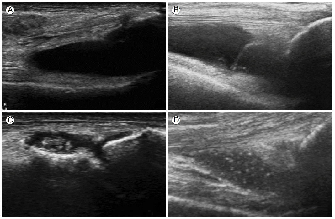
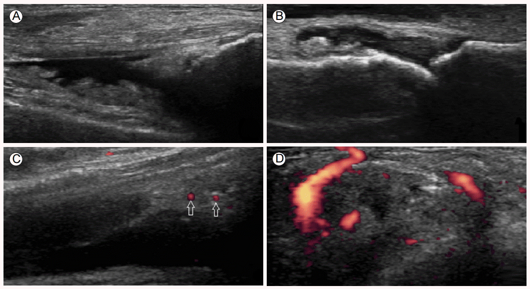
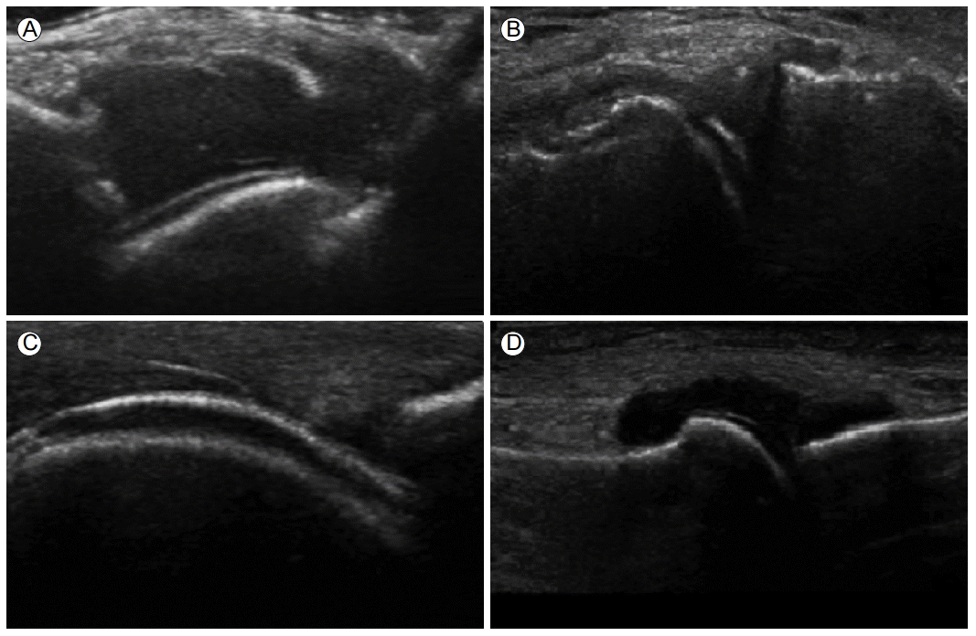
ÝÖ£ýòíýØÿ ýªØÛ░ÇÙèö Û▓░ýáòý£áÙ░£ýä▒ Û┤Çýáêýù╝ýùÉ Ýè╣ýØ┤ýáüýØ© ýåîÛ▓¼ýØÇ ýòäÙïêÙ®░, ÝåÁÝÆìýØ┤Ùéÿ CPPD ý╣¿ý░®ýºêÝÖÿ Ù¬¿ÙæÉýùÉýä£ ÝØöÝ×ê Û┤Çý░░ÙÉ£Ùïñ. Û▓░ýáòý£áÙ░£ýä▒ Û┤Çýáêýù╝ýØÿ ý┤êÛ©░ýùÉÙèö Ù¼┤ýùÉý¢ö ýØîýÿüýØÿ ýØ╝Ù░ÿ Û┤Çýáêýòí ýûæýâüý£╝Ùí£ Û┤Çý░░ÙÉá ýêÿ ý×êý£╝Ùéÿ(Fig. 1A and 1B), ýï£Û░äýØ┤ ýºÇÙéÿÙ®┤ýä£ Ùé┤ÙÂÇýùÉ ÙïñýûæÝò£ ýùÉý¢ö ýØîýÿüýØä ÙéÿÝâÇÙé┤Ùèö ýÜöýé░ Û▓░ýáòýØÿ ýØæýºæýØä Ù│┤ýØ┤Û▓î ÙÉ£Ùïñ(Fig. 1C) [3,16-18]. 1 mm ýØ┤ÝòÿýØÿ Û│áýùÉý¢ö ýØîýÿüýØÿ ýØæýºæý▓┤ÙôñýØ┤ ÔÇ£starry skyÔÇØ ýºòÝøäÙí£ Û┤Çý░░ÙÉÿÛ▒░Ùéÿ (Fig. 1D) [19], ÝâÉý┤ëý×ÉÙí£ ýáüýáêÝò£ ýòòÙ░òýØä Û░ÇÝòÿÛ▓î ÙÉÿÙ®┤ ýØ┤Ùƒ¼Ýò£ ýØæýºæý▓┤ÙôñýØ┤ Û┤Çýáê Ùé┤Û░òý£╝Ùí£ ÙûáýÿñÙÑ┤Ù®░ ÔÇ£snow-stormÔÇØ ÝÿòýâüýØä ÙéÿÝâÇÙé┤Û©░ÙÅä Ýò£Ùïñ[12,18,20]. ýØ┤Ùƒ░ ýØæýºæý▓┤ÙôñýØÇ Û┤ÇýáêÛ░ò Ùé┤ÙÂÇýØÿ Û▒░ÝÆêýØ┤Ùéÿ ÝîîÝÄ© Û░ÖýØÇ Ù╣äÛ▓░ýáòýä▒ Û│áýùÉý¢ö ýØîýÿü Ù¼╝ýºêÙôñÛ│╝ ÛÁ¼ÙÂäÝòÿýù¼ýò╝ ÝòÿÙèöÙì░[12,21], ý┤êýØîÝîî Û©░Û©░ýØÿ ýªØÝÅ¡(gain)ýØä Ùé«ýÂöÙèö Û▓âýØ┤ ÙÅäýøÇýØ┤ ÙÉ£Ùïñ[22]. ýØ┤Ùƒ¼Ýò£ ÝÖ£ýòí Ùé┤ÙÂÇýùÉýä£ Û┤Çý░░ Û░ÇÙèÑÝò£ Û│áýùÉý¢ö ýØîýÿüýØÿ ýØæýºæý▓┤ÙôñýØÇ Ýè╣ýáò Û▓░ýáòý£áÙ░£ýä▒ Û┤Çýáêýù╝ýùÉ Ýè╣ýØ┤ýáüýØ© ýåîÛ▓¼ýØÇ ýòäÙïêýºÇÙºî[4,20], Û▓░ýáòý£áÙ░£ýä▒ Û┤Çýáêýù╝Û│╝ Ù╣äÛ▓░ýáò Û┤Çýáêýù╝ýØÿ ÛÁ¼ÙÂäýùÉÙèö ÙÅäýøÇýØ┤ ÙÉá ýêÿ ý×êÙïñ.
ÝÖ£ÙºëýØÿ ýªØýïØ(synovial hypertrophy or proliferation)
ÝÖ£ÙºëýØÿ ýªØýïØ ýù¡ýï£ Û▓░ýáòý£áÙ░£ýä▒ Û┤Çýáêýù╝ýùÉýä£ Û┤Çý░░ÙÉá ýêÿ ý×êÙèö Ù╣äÝè╣ýØ┤ýáüýØ© ýåîÛ▓¼ý£╝Ùí£, ÙæÉÝä░ýøîýºä ÝÖ£Ùºë Ù▓¢ýØÿ Ù╣äÝøäÙí£ Û┤Çý░░ÙÉÿÙ®░(Fig. 2A and 2B), Ýîîýøî ÙÅäÝöîÙƒ¼Ùí£ ÝÿêÙÑÿýØÿ ýªØÛ░ÇÛ░Ç ÙÅÖÙ░ÿÙÉ£ Û▓âýØä ÝØöÝ×ê Ù│╝ ýêÿ ý×êÙïñ(Fig. 2C and 2D). ÝÿêÙÑÿýØÿ ýªØÛ░ÇÛ░Ç grade 2-3 (0 = no vessel in the synovium; 1 = up to 3 single spots signals or 1 confluent spot + up to 2 single spots; 2 = vessel signals in less than half of the area of the synovium; 3 = vessel signals in more than half of the area of the synovium) ýáòÙÅäÙí£ ýªØÛ░ÇÙÉÿýû┤ ý×êÙïñÙ®┤, ýâêÙí£ýØ┤ Ù░£ýâØÝò£ Û©ëýä▒ Ù│æÙ│ÇýØ╝ Û░ÇÙèÑýä▒ýØ┤ Ýü¼Ùïñ[22]. ý╣ÿÙúîÛ░Ç ý×ÿ ýØ┤Ùú¿ýû┤ýºê Û▓¢ýÜ░ ýÂöýáü Û▓Çýé¼ýùÉýä£ ÙÅäÝöîÙƒ¼ ýØîýÿüýØÿ Û░Éýåî Ù░Å ýåîýïñýØä Û┤Çý░░Ýòá ýêÿ ý×êÙèöÙì░[23], ýØ┤Ùƒ¼Ýò£ ýåîÛ▓¼ýØÇ ý╣ÿÙúî ÝÜ¿Û│╝ ÝÅëÛ░ÇýùÉ Û░äÝÄ©ÝòÿÛ│á ý£áýÜ®ÝòÿÛ▓î ýô░ýØ╝ ýêÿ ý×êÙïñ[24].
ÝåÁÝÆìýä▒ Û┤Çýáêýù╝
ýÜöýé░ ýØæýºæý▓┤(MSU aggregates)
Ýÿêýñæ ýÜöýé░ ýêÿý╣ÿÛ░Ç 6.8 mg/dLýØä ý┤êÛ│╝ÝòÿÛ▓î ÙÉÿÙ®┤ ÝÿêýòíÙé┤ Û│╝ÝżÝÖö ýâüÝâ£Û░Ç ÙÉÿýû┤ ý▓┤Ùé┤ýùÉ Ùà╣ýòäý×êÙìÿ ýÜöýé░ýØ┤ Û▓░ýáòÝÖöÙÉ£Ùïñ[25]. ýØ┤Ùíáýáüý£╝Ùí£Ùèö ýâØý▓┤Ùé┤ Ù¬¿Ùôá ÛÁ¼ýí░Ù¼╝ýùÉ ý╣¿ý░®ýØ┤ Û░ÇÙèÑÝòÿÙ®░, ýú╝Ù│Ç ýí░ýºüýùÉ Ù╣äÝò┤ ý┤êýØîÝîîÙÑ╝ Û░òÝòÿÛ▓î Ù░ÿýé¼ÝòÿÛ©░ ÙòîÙ¼©ýùÉ ÛÁ¼Ù│äýØ┤ ýë¢Û│á, ÝâÉý┤ëý×ÉýØÿ Û░üÙÅäýùÉ ýÿüÝûÑýØä ýáüÛ▓î Ù░øÙèöÙïñ[4,12,20,26]. ýÜöýé░ Û▓░ýáòýØÿ ý╣¿ý░®Ù¼╝ýØÇ ýºêÙ│æýØÿ Ù│æÛ©░, ý╣¿ý░®ÙÉ£ ÙÂÇý£ä, ÙÂäÝż, Ýü¼Û©░ýùÉ Ùö░ÙØ╝ ý┤êýØîÝîî ýâüýùÉýä£ Û░üÛ░ü ÙïñÙÑ© Ýè╣ýºòýáüýØ© ýûæýâüýØä Ù│┤ýØ┤Û▓î ÙÉ£Ùïñ[26].
Hyperechoic spot
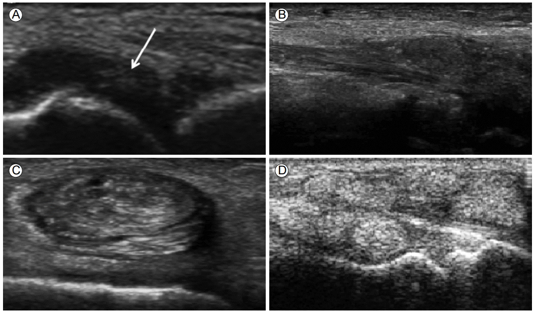
1 mm ýØ┤ÝòÿýØÿ ýÜöýé░ Û▓░ýáòý£╝Ùí£ ÝåÁÝÆì ÝÖÿý×ÉýØÿ ý┤êýØîÝîî Û▓Çýé¼ýï£ Û░Çý×Ñ ÝØöÝòÿÛ▓î Û┤Çý░░ÙÉÿÙèö ýåîÛ▓¼ýØ┤Ùïñ. Û┤Çýáêýòí ýòêýùÉýä£ Û┤Çý░░ÙÉá Û▓¢ýÜ░ ÔÇÿÙêêÙ│┤ÙØ╝(snow storm)ÔÇÖ ýºòÝøäÙÑ╝ Ù│┤ýØ┤Ù®░, ÝÖ£Ùºë ýªØýïØ ÙÂÇý£ä ýòêýùÉýä£ Û┤Çý░░ÙÉÿÙèö Û▓âýØÇ ÝåÁÝÆìýùÉ Ýè╣ýØ┤ýáüýØ© ýåîÛ▓¼ý£╝Ùí£ ýâØÛ░üÙÉÿýû┤ýºÇÛ│á ý×êÙïñ[22].
Hyperechoic cloudy area
1 cm ýØ┤ÝòÿýØÿ ýÜöýé░ Û▓░ýáòýØÿ ýØæýºæý▓┤Ùí£, ýØ╝Ù░ÿýáüý£╝Ùí£ ÝøäÙ░® ýØîýÿü Û░ÉýåîÙÑ╝ ÙÅÖÙ░ÿÝòÿýºÇ ýòèÙèö ÛÀáýØ╝Ýò£ ýûæýâüýØÿ ýØîýÿüýØä Ù│┤ýØ©Ùïñ. ýú╝ý£ä ýí░ýºüÛ│╝ Ù¬àÝÖòÝòÿÛ▓î ÛÁ¼ÙÂäÙÉÿÙèö ÝÄ©ýØ┤ýºÇÙºî ÛÀ©ÙáçýºÇ ýòèýØÇ Û▓¢ýÜ░ÙÅä ý×êýû┤ Ùºêý╣ÿ ÛÁ¼Ùªäý▓ÿÙƒ╝ Ù│┤ýØ┤Ùèö ýåîÛ▓¼ýØ┤Ùïñ(Fig. 3A) [27]. ý╣ÿÙúîÙí£ ýë¢Û▓î ýåîýïñÙÉÿÙèö Ýè╣ýºòýØ┤ ý×êÙïñ[28].
ýÜöýé░ Û▓░ýáê(Tophi)
ýä©Ýż Ù░ûýùÉ ýÜöýé░ Û▓░ýáòýØ┤ ý╣¿ý░®ÝòÿÛ▓î ÙÉÿÙ®┤, ýØ┤Ù¼╝ Û▒░ÙîÇýä©Ýż(foreign body giant cell)ýÖÇ Ùï¿ÝòÁÛÁ¼ÙôñýùÉ ÙæÿÙƒ¼ýï©ýù¼ ý£íýòäýóàÛ│╝ Ù╣äýèÀÝò£ ÛÁ¼ýí░Ù¼╝ýØä ÙºîÙôñÛ▓î ÙÉÿÙèöÙì░, ýØ┤ÙÑ╝ ýÜöýé░ Û▓░ýáêýØ┤ÙØ╝ Ýò£Ùïñ[29]. ýû┤ÙèÉ ÙÂÇý£äýùÉýä£Ùéÿ Ù░£ýâØýØ┤ Û░ÇÙèÑÝòÿÙéÿ ýú╝Ùí£ Ù░£Û░ÇÙØ¢(Ýè╣Ý×ê ý▓½ýº© Ù░£ÝùêÙª¼Ù░£Û░ÇÙØ¢ Û┤Çýáê), ýåÉÙ¬®, ýåÉÛ░ÇÙØ¢, Ù¼┤ÙªÄ, Ù░£Ù¬® Ùô▒ýùÉýä£ Û┤Çý░░ÙÉ£Ùïñ[3,19]. ýòòýÂòÙÉ£ ýáòÙÅäýùÉ Ùö░ÙØ╝ ÙïñýûæÝò£ ý┤êýØîÝîî ýÿüýâüýåîÛ▓¼ýØä ÙéÿÝâÇÙé┤ÙèöÙì░ soft, hard, mixedýØÿ ýä© Û░ÇýºÇ ýóàÙÑÿÙí£ ÙÂäÙÑÿÙÉ£Ùïñ[12,18,20,30]. Soft tophiÙèö ý┤ëýºÇýï£ ÙÂÇÙô£Ùƒ¢Û│á ýáÇýùÉý¢ö ýØîýÿüýØÿ ý×æÛ│á ÛÀáýØ╝Ýò£ Û▓░ýáêÙí£ Û┤Çý░░ÙÉÿÙ®░, Û│áýùÉý¢ö ýØîýÿüýØä ÙØäÙèö ÝàîÙæÉÙª¼ýùÉ ÙæÿÙƒ¼ýï©ýØ© ýûæýâüýØä Ù│┤ýØ┤Û©░ÙÅä Ýò£Ùïñ(Fig. 3B) [5]. Û▓░ýáòýØ┤ ýºÇýåìýáüý£╝Ùí£ ý╣¿ý░®ÝòÿÛ▓î ÙÉÿÙ®┤ ýáÉý░¿ Û│áýùÉý¢ö ýØîýÿüýØÿ Û©░ýºêÙí£ Ù│ÇÝÖöÝòÿÙ®┤ýä£ ýú╝Ù│Ç ýí░ýºüÛ│╝ ÝÖòýù░Ý×ê ÛÁ¼ÙÂäÙÉÿÙ®░, Ýü¼Û©░Û░Ç ý╗ñýá© ýú╝ý£ä ýí░ýºüÙôñýØä ýòòÙ░òÝòÿÛ▓î ÙÉ£Ùïñ. ýêÿÙàäýØ┤ Û▓¢Û│╝ÝòÿÛ▓î ÙÉÿÙ®┤ ý┤ëýºÇýï£ Ùï¿Ùï¿ÝòÿÛ│á, ÝøäÙ░® ýØîýÿü Û░ÉýåîÙÑ╝ ÙÅÖÙ░ÿÝòÿÙèö ÙÂêÛÀáýºêÝò£ ýØîýÿüýØÿ ýóàÛ┤┤Ùí£ Ù░£Ùï¼ÝòÿÛ▓î ÙÉÿÙèöÙì░ ýØ┤ÙÑ╝ hard ÙÿÉÙèö mixed tophiÙí£ Ýæ£ÝÿäÝò£Ùïñ(Fig. 3C and 3D). ýØ┤ Ùï¿Û│äýØÿ tophiÙèö ÙîÇýïØ ýä©ÝżÙéÿ ý×äÝîîÛÁ¼, Û▒░ÙîÇýä©ÝżÙôñýùÉ ýØÿÝò£ ýù╝ýªØ Ù░ÿýØæýØä ý£áÙ░£Ýòÿýù¼ ýáÇýùÉý¢ö ýØîýÿüýØÿ ÝàîÙæÉÙª¼ÙÑ╝ ÙÅÖÙ░ÿÝòÿÛ©░ÙÅä ÝòÿÙ®░[31], ýù╝ýªØ Ù░ÿýØæýØä ÝåÁÝò┤ ýØ©ýáæÝò£ Ù╝êýØÿ Ù»©Ù×ÇÛ│╝ ÝîîÛ┤┤ÙÑ╝ ý£áÙ░£Ýò£Ùïñ[9]. ýªØýâüýØ┤ ýùåÙèö ýÜöýé░ Û▓░ýáêýä▒ ÝåÁÝÆì ÝÖÿý×ÉýùÉýä£ Û│¿Ù»©Ù×ÇýØ┤ ýºÇýåìýáüý£╝Ùí£ ýºäÝûëÝòÿÙèö Û▓âýØÇ ýØ┤Ùƒ¼Ýò£ ýÜöýé░ Û▓░ýáêýùÉ ýØÿÝò£ Ùºîýä▒ýáüýØ© ýù╝ýªØ Ù░ÿýØæýùÉýä£ Û©░ýØ©Ýòá Û▓âý£╝Ùí£ ýâØÛ░üÙÉÿýû┤ýºÇÛ│á ý×êÙïñ[32].
Û▒┤Ùé┤ ýÜöýé░ Û▓░ýáêýØÇ ýè¼Û░£Û│¿ ýØ©ÙîÇ(patellar ligament), ýòäÝé¼ÙáêýèñÛ▒┤(achillestendon), ýé╝ÙæÉÛÀ╝Û▒┤(tricepstendon), ÙîÇÝç┤ýé¼ÙæÉÛÀ╝Û▒┤(quadricipital tendon), ýò×ýáòÛ░òÛÀ╝Û▒┤(tibialis anterior tendon), Ù░£Ù░öÙïÑÛÀ╝Ùºë(plantar fascia)ýùÉýä£ ý×ÿ Û┤Çý░░ÙÉÿÙèöÙì░, Û▓¢Û│äÛ░Ç Ù¬àÝÖòÝòÿýºÇ ýòèÛ│á ÙÂêÛÀáýºêÝò£ ýûæýâüýØÿ Û│á/ýáÇýùÉý¢ö ýØîýÿüýØÿ Ù│æÙ│Çý£╝Ùí£ Ù│┤ýØ┤Ù®░, ÝøäÙ░® ýØîýÿüÛ░ÉýåîÙÑ╝ ÙÅÖÙ░ÿÝòÿÛ©░ÙÅä Ýò£Ùïñ. ýØ┤Ùƒ¼Ýò£ ÝåÁÝÆì Û▓░ýáêýØÇ Û▒┤ýØä ýò¢ÝÖöýï£ý╝£ ýë¢Û▓î ýåÉýâüýØä ý£áÙ░£Ýòá ýêÿ ý×êÙïñ[33].
ÛÀ© ýÖ©ýùÉ Û┤Çýáê ýú╝ý£ä ýáÉýòíÙé¡ýØÿ Û▓¢ýÜ░ ýáäýè¼Û░£ÝÖ£ýòíÙé¡(prepatellar bursa), ý▓ÖÛ│¿ÙæÉÝÖ£ýòíÙé¡(olecrenon bursa)ýùÉýä£ Û░Çý×Ñ ý×ÿ Û┤Çý░░ÙÉÿÙ®░[34,35], Ýö╝Ýòÿ ýí░ýºüÙé┤ ÝåÁÝÆì Û▓░ýáêýØÿ Û▓¢ýÜ░ ÙÑÿÙºêÝï░ýèñÛ▓░ýáê Ùô▒ ÙïñÙÑ© Ù│æÙ│ÇÛ│╝ Û░ÉÙ│äÝò┤ýò╝ ÝòÿÙèöÙì░, 80%ýùÉýä£ ÙÂêÛÀáýºêÝòÿÛ│á Û│áýùÉý¢ö ýØîýÿüýØÿ ýåîÛ▓¼ýØä Ù│┤ýØ┤Û©░ ÙòîÙ¼©ýùÉ Û░ÉÙ│äýØ┤ Û░ÇÙèÑÝòÿÙïñ[36,37].
ýØ┤Ùƒ¼Ýò£ ÝåÁÝÆìýùÉ Ýè╣ýØ┤ýáüýØ© ýÜöýé░ Û▓░ýáêýØÇ MRIÙéÿ DECT Ùô▒ ÙïñÙÑ© ýÿüýâüÝòÖýáü Û▓Çýé¼Ùí£ÙÅä ýºäÙï¿ýØ┤ Û░ÇÙèÑÝòÿÙïñ[38]. ÛÀ©Ùƒ¼Ùéÿ ýÜöýé░ ýêÿý╣ÿÙÑ╝ ýáüýáêÝòÿÛ▓î ý£áýºÇÝòá Û▓¢ýÜ░ ýÜöýé░ Û▓░ýáêýØÿ ýºüÛ▓¢Û│╝ ÙÂÇÝö╝Û░Ç Û░ÉýåîÝò¿ýØ┤ ÝÖòýØ©ÙÉ¿ýùÉ Ùö░ÙØ╝[26], ýØ┤ÙÑ╝ ý©íýáòÝòÿÛ©░ ýÜ®ýØ┤Ýò£ ý┤êýØîÝîî ýÿüýâü Û▓Çýé¼Û░Ç ýºêÙ│æýØÿ ý╣ÿÙúî ÝÜ¿Û│╝ ÝîÉýáòýùÉ Ùìö ý£áÙª¼ÝòÿÛ©░ ÙòîÙ¼©ýùÉ ÝåÁÝÆìýØÿ ýÂöýáüýºäÙúîýùÉ Ù│┤Ùïñ ýáüÝò®Ýò£ Û▓Çýé¼ Ù░®Ù▓òý£╝Ùí£ýä£ ÙäÉÙª¼ ýØ┤ýÜ®ÙÉÿÛ│á ý×êÙïñ.
Û│¿Ù»©Ù×Ç(bone erosion)
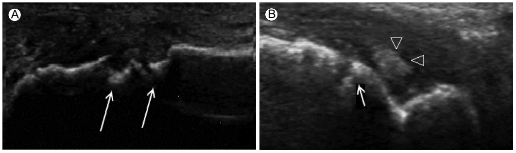
Û│¿Ù»©Ù×ÇýØÇ ýºüÛ░üýØÿ ÙæÉ ÝâÉý┤ë Ù®┤ýùÉýä£ Ù¬¿ÙæÉ Û┤Çý░░ÙÉÿÙèö Û│¿Ýö╝ýºêýØÿ Û▓░ýåÉý£╝Ùí£, ÝåÁÝÆìýùÉ ýØ┤ÝÖÿÙÉ£ Û©░Û░äýØ┤ Û©©Û│á Ù░£ý×æ Ý܃ýêÿÛ░Ç ÙºÄÛ▒░Ùéÿ, ÝåÁÝÆì Û▓░ýáêýØ┤ ÙÅÖÙ░ÿÙÉÿýû┤ ý×êÙèö ýºêÝÖÿýØÿ Û▓¢ýÜ░ýùÉ Ùìö ýë¢Û▓î Û┤Çý░░ÙÉá ýêÿ ý×êÙïñ(Fig. 4A and 4B) [39]. ÝåÁÝÆìýùÉ ýØÿÝò£ Û│¿Ù»©Ù×ÇýØ┤ ÝØöÝ×ê Û┤Çý░░ÙÉÿÙèö ÙÂÇý£äÙèö ý▓½ýº© Ù░£ÝùêÙª¼Ù░£Û░ÇÙØ¢Û┤ÇýáêýØÿ Ùé┤ý©íÛ│╝ ýåÉÝùêÙª¼ýåÉÛ░ÇÙØ¢Û┤ÇýáêýØ┤Ùïñ[19,24,40]. ý▓½ýº© Ù░£ÝùêÙª¼Ù░£Û░ÇÙØ¢Û┤ÇýáêýùÉýä£ Û│¿Ù»©Ù×ÇýØ┤ Ù░£Û▓¼ÙÉÿÙèö Ù╣êÙÅäÙèö ýáòýâü ÙîÇýí░ÛÁ░ýùÉýä£ 6%, ÙïñÙÑ© Û┤Çýáêýù╝ýùÉ ýØ┤ÝÖÿÙÉ£ ÝÖÿý×ÉÛÁ░ýùÉýä£ 43%ÙÑ╝ Ù│┤ýØ© Ùì░ýùÉ Ù╣äÝò┤, ÝåÁÝÆì ÝÖÿý×ÉýùÉýä£Ùèö 67%Ùí£ ÙåÆÛ▓î ÙéÿÝâÇÙé£Ùïñ[39].
ÝåÁÝÆìýùÉ ýØÿÝò£ Û│¿Ù»©Ù×ÇýØÿ Û▓¢ýÜ░, ÙÑÿÙºêÝï░ýèñÛ┤Çýáêýù╝ Ùô▒ ÙïñÙÑ© Ùºîýä▒ Û┤Çýáêýù╝ýùÉ ýØÿÝò£ Û│¿Ù»©Ù×ÇýùÉ Ù╣äÝò┤ Ùìö Û╣èÛ│á, ÝîîÛ┤┤ýáüýØ© ýûæýâüýØä Ù│┤ýØ┤Ù®░, Ù╣äÙîÇý╣¡ýáüý£╝Ùí£ Ù░£ýâØÝòÿÛ│á, ýú╝ý£ä Ù╝êýØÿ Û▓¢ÝÖöÙÑ╝ ÙÅÖÙ░ÿÝò£ÙïñÙèö ýáÉýùÉýä£ ý░¿ýØ┤Û░Ç ý×êý£╝Ùéÿ, Ùï¿ÙÅàý£╝Ùí£Ùèö ÝåÁÝÆìýØÿ ýºäÙï¿ýùÉ Ýè╣ýØ┤ýáüýØ┤ýºÇ ýòèý£╝Ù®░, Û│¿Ù»©Ù×Ç ýú╝ý£äýØÿ ýÜöýé░ Û▓░ýáò ý╣¿ý░®ýØ┤Ùéÿ ýÜöýé░ Û▓░ýáêýØÿ ÙÅÖÙ░ÿ ýù¼ÙÂÇÙÑ╝ ÝÖòýØ©Ýòÿýù¼ýò╝ Ýò£Ùïñ[18,36].
Ýÿäý×¼Ùí£ýä£Ùèö ÝåÁÝÆìýùÉ ýØÿÝò£ Û│¿Ù»©Ù×ÇýØÿ Ùô▒Û©ë Û©░ýñÇ(scoring system)ýØ┤ ýáòÙª¢ÙÉÿýºÇ ýòèýòä Wakefield Ùô▒[41]ýØ┤ ÙÑÿÙºêÝï░ýèñÛ┤Çýáêýù╝ýØÿ Û│¿Ù»©Ù×ÇýùÉýä£ ýá£ýï£Ýò£ ýáòÙƒëýáü Û©░ýñÇ(small < 2 mm, moderate 2-4 mm, large erosions > 4 mm)ýØä ýØ╝Ù░ÿýáüý£╝Ùí£ ýé¼ýÜ®ÝòÿÛ│á ý×êÙïñ.
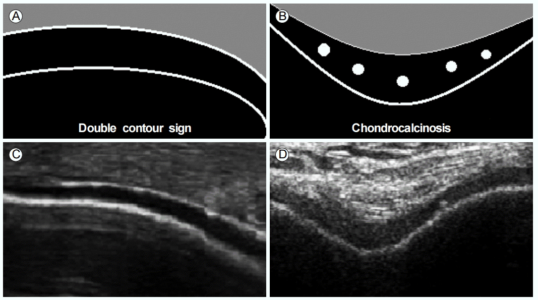
Û┤Çýáê ýù░Û│¿ýØÿ Ýè╣ýºòýáüýØ© ýÜöýé░ Û▓░ýáòýØÿ ý╣¿ý░®
ýáòýâü ý£áÙª¼ýºê ýù░Û│¿ýØÇ ý┤êýØîÝîîýùÉýä£ Û│áýùÉý¢ö ýØîýÿüýØä ÙØäÙèö ÙæÉ Û░£ýØÿ ýäáÙ¬àÝò£ Û▓¢Û│äÙ®┤ý£╝Ùí£ ÙæÿÙƒ¼ýï©ýù¼ ýú╝Ù│Ç ýí░ýºüÛ│╝ Ù¬àÝÖòÝ×ê ÛÁ¼ÙÂäÙÉÿÙèö ÛÀáýºêÝò£ ýûæýâüýØÿ Ù¼┤ýùÉý¢ö ýØîýÿü ý©Áý£╝Ùí£ Û┤Çý░░ÙÉ£Ùïñ. ýù░Û│¿ýØÿ Ýæ£ý©ÁÙ®┤ýØÇ ÝâÉý┤ëý×ÉýØÿ Û░üÙÅäÙÑ╝ ýêÿýºüý£╝Ùí£ ýí░ýáêÝòÿÙ®┤ Û┤Çý░░ýØ┤ ýÜ®ýØ┤Ýò┤ýºÇÙèöÙì░, ýáòýâüýáüý£╝Ùí£ Ýæ£ý©ÁýØÿ Û▓¢Û│äÙ®┤ýØ┤ ýï¼ÙÂÇýØÿ Û▓¢Û│äÙ®┤Ù│┤Ùïñ ýûçÛ▓î ÙéÿÝâÇÙé£Ùïñ. ýÜöýé░ Û▓░ýáòýØÇ ýù░Û│¿ýØÿ Ýæ£ý©ÁÙ®┤ýØä ýäáÝÿ©ÝòÿÙèö Û▓¢ÝûÑýØ┤ ý×êýû┤ ýú╝Ùí£ ýØ┤ ÙÂÇý£äýùÉ ý╣¿ý░®ÙÉÿÙ®░[22], ýØ┤Ùí£ ýØ©Ýò┤ ý╣¿ý░®ÙÉ£ ýù░Û│¿ýØÿ Ýæ£ý©ÁÙ®┤ýØÇ ýÜöýé░ Û▓░ýáòýØÿ ý╣¿ý░® ýáòÙÅäýùÉ Ùö░ÙØ╝ ÛÁ¡ýåîýáüý£╝Ùí£, Ýÿ╣ýØÇ Ù»©Ùºîýä▒ý£╝Ùí£ ýù░Û│¿Ýòÿ Û│¿Ýö╝ýºêýùÉ ÝòäýáüÝòÿÙèö Û│áýùÉý¢ö ýØîýÿüýØä ÙØäÛ▓î ÙÉ£Ùïñ. ýØ┤Û▓âýØä ÔÇÿýØ┤ýñæý£ñÛ│¢(double contour, DC) ýºòÝøäÔÇÖÙØ╝Û│á ÝòÿÙ®░ ÙîÇÛ░£ ÝâÉý┤ëý×ÉýØÿ Û░üÙÅäýÖÇ ýâüÛ┤ÇýùåýØ┤ ÙÜ£ÙáÀÝòÿÛ▓î Û┤Çý░░ÙÉ£Ùïñ(Fig. 5) [9,12,25,39]. DC ýºòÝøäÙèö ÝåÁÝÆì ýºäÙï¿ýùÉ ý×êýû┤ýä£ Ù»╝Û░ÉÙÅäÙèö 43.6%Ùí£ Ùïñýåî Ùé«ýØÇ Ù░ÿÙ®┤, Ýè╣ýØ┤ÙÅäÙèö 99%Ùí£ ÙºñýÜ░ ÙåÆÛ│á[25], ÙïñÙÑ© Û┤Çýáê ýºêÝÖÿýùÉýä£Ùèö ÙéÿÝâÇÙéÿýºÇ ýòèÛ│á ýÿñýºü ÝåÁÝÆìýùÉýä£Ùºî Û┤Çý░░ÙÉÿÛ©░ ÙòîÙ¼©ýùÉ Û░ÉÙ│ä ýºäÙï¿ýùÉ ý£áýÜ®ÝòÿÙïñ[39]. ÙÿÉÝò£ ý┤êýØîÝîî ýêáý×É Û░ä ýêÖÙá¿ÙÅäýØÿ ý░¿ýØ┤ýùÉÙÅä ÙÂêÛÁ¼ÝòÿÛ│á Û▓Çýé¼ýØÿ ý×¼ýù░ýä▒ýØ┤ ÙåÆÙïñÙèö Ýè╣ýºòýØ┤ ý×êýû┤[25,42], ÝåÁÝÆì ýºäÙï¿ýùÉ ý×êýû┤ Û░Çý×Ñ ýñæýÜöÝòÿÛ│á, ýïáÙó░ÝòáÙºîÝò£ ýåîÛ▓¼ ýñæ ÝòÿÙéÿÙí£ Û╝¢Ý×êÛ│á ý×êÙïñ. DC ýºòÝøäÙèö Ù░£ÝùêÙª¼Ù░£Û░ÇÙØ¢, Ù¼┤ÙªÄ, ýåÉÝùêÙª¼ýåÉÛ░ÇÙØ¢Û┤ÇýáêýùÉýä£ ý×ÿ Û┤Çý░░ÙÉÿÙ®░, Û│¿Û┤Çýáêýù╝ýùÉ ýØ┤ÝÖÿÙÉ£ Û┤Çýáêý▓ÿÙƒ╝ ýù░Û│¿ýØ┤ ýûçýØÇ Û▓¢ýÜ░ýùÉýä£Ùèö ý×ÿ Û┤Çý░░ÙÉÿýºÇ ýòèÙèöÙïñ[4]. ÙÿÉÝò£, DC ýºòÝøäÙèö CPPD Û▓░ýáòýØ┤ ýù░Û│¿ Ùé┤ÙÂÇýùÉ ÙØá Ù¬¿ýûæý£╝Ùí£ ý╣¿ý░®ÙÉÿÛ▒░Ùéÿ, ýªØÛ░ÇÙÉ£ Û┤Çýáêýòíý£╝Ùí£ ýØ©Ýò£ ÝøäÙ░® ýí░ýÿü ýªØÛ░ò ÝÜ¿Û│╝Ùí£ ýØ©Ýò┤ ýáòýâü ýù░Û│¿ýØÿ Ýæ£ý©ÁÙ®┤ýØ┤ Û│áýùÉý¢ö ýØîýÿüý£╝Ùí£ Û┤Çý░░ÙÉÿÙèö Û▓¢ýÜ░ýÖÇ Ù░ÿÙô£ýï£ Û░ÉÙ│äÝòÿýù¼ýò╝ Ýò£Ùïñ[4,12,20].
Ù¼┤ýªØýâü Û│áýÜöýé░ÝÿêýªØ ÝÖÿý×É 26Ù¬àýØä ÙîÇýâüý£╝Ùí£ Ýò£ ý┤êýØîÝîî ýù░ÛÁ¼ýùÉýä£ 27%ýùÉ Ýò┤Ùï╣ÝòÿÙèö 7Ù¬àýØÿ ÝÖÿý×ÉýùÉýä£ DC ýºòÝøäÛ░Ç Û┤Çý░░ÙÉÿýùêÙèöÙì░, ýØ┤Ùèö DC ýºòÝøäÛ░Ç ýºêÙ│æýØÿ Ù│æÛ©░ýÖÇÙèö Ýü░ Û┤ÇÙá¿ýØ┤ ýùåÙïñÙèö Û▓âýØä Ù│┤ýù¼ýñÇÙïñ[43]. ÛÀ©Ùƒ¼Ùéÿ ýªØýâüýØ┤ ýùåÙìöÙØ╝ÙÅä ýÜöýé░ Û▓░ýáòýØÿ ý╣¿ý░®ýØ┤ Û┤Çý░░ÙÉÿÙèö Û▓¢ýÜ░, ÝûÑÝøä ÝåÁÝÆìýä▒ Û┤Çýáêýù╝ýØÿ Ù░£ýâØ Û░ÇÙèÑýä▒ýØ┤ ÙåÆýòäýºÇÛ©░ ÙòîÙ¼©ýùÉ ýØ┤ ýï£Û©░ÙÂÇÝä░ ÝåÁÝÆìý£╝Ùí£ ýºäÙï¿Ýò┤ýò╝Ýò£ÙïñÙèö ýØÿÛ▓¼ýØ┤ ÙºÄýòäýºÇÛ│á ý×êÙïñ. ÙÿÉÝò£ DC ýºòÝøäÛ░Ç Û┤Çý░░ÙÉÿÙèö ÝåÁÝÆì ÝÖÿý×ÉýùÉýä£ ýÜöýé░ýØÿ ÙåìÙÅäÛ░Ç ýûæýØÿ ýâüÛ┤ÇÛ┤ÇÛ│äÙÑ╝ Ù│┤ýØ©ÙïñÙèö ýáÉýùÉýä£[44] ýºêÙ│æýØÿ Û▓¢ýñæýØä ÙîÇÙ│ÇÝòÿÙèö ýºÇÝæ£Ùí£ ýù¼Û▓¿ýºÇÛ│á ý×êý£╝Ù®░[45], ÝåÁÝÆìýØÿ ý╣ÿÙúîÛ░Ç ý×ÿ ýØ┤Ùú¿ýû┤ýºê Û▓¢ýÜ░ ýåîýïñÙÉÿÙèö Ýè╣ýä▒ýØä ýØ┤ýÜ®Ýòÿýù¼ ý╣ÿÙúîýØÿ ÝÜ¿Û│╝ÙÑ╝ ÝÅëÛ░ÇÝòÿÙèö Ùì░ÙÅä ÝÖ£ýÜ®Ýòá ýêÿ ý×êÙïñ[28,38,46].
CPPD ý╣¿ý░®ýºêÝÖÿ
CPPD ý╣¿ý░®ýºêÝÖÿýØÇ Û│áÙá╣ ýØ©ÛÁ¼ýùÉýä£ ÝØöÝò£ Û▓░ýáò ý╣¿ý░®ýºêÝÖÿý£╝Ùí£ 65-75ýä©ýùÉýä£ 10-15%, 85ýä© ýØ┤ýâüýùÉýä£ 30-60%Ùí£ Ù░£ýâØÝò£Ùïñ. ÝÖÿý×ÉýØÿ 80% ýØ┤ýâüýØ┤ 60ýä© ýØ┤ýâüýØ┤Û│á 70% ýØ┤ýâüýØÿ Û▓¢ýÜ░ýùÉýä£ Û│╝Û▒░ Û┤Çýáê ýåÉýâüýØ┤ ý×êýùêÙìÿ Û▓âý£╝Ùí£ Ù│┤ýòä Ùà©ÝÖöÙÉ£ ýù░Û│¿ýØÿ ýâØÝÖöÝòÖýáüýØ© Ù│ÇÝÖöÛ░Ç Û▓░ýáò ý╣¿ý░®ýùÉ Û┤Çýù¼ÝòÿÙèö Û▓âý£╝Ùí£ ýâØÛ░üÙÉ£Ùïñ. Ýè╣Ý×ê Û│¿Û┤Çýáêýù╝ýØ┤ ý×êÛ▒░Ùéÿ Ù¼┤Û©░Ýö╝Ùí£ýØ©ýé░ýù╝(inorganic pyrophosphate)ýØ┤ Û┤ÇýáêÛ░ò Ùé┤ýùÉ ÙºÄýØÇ Û▓¢ýÜ░, ÙîÇýé¼ ýºêÝÖÿýØ┤ ÙÅÖÙ░ÿÙÉ£ Û▓¢ýÜ░ýùÉ Û┤ÇýáêÙé┤ Û▓░ý▓┤ ýí░ýºü ÙÂÇý£ä ýñæ ÝÿêÛ┤ÇýØ┤ ýùåÙèö ýù░Û│¿ ÙÂÇý£äýùÉ ý╣╝ýèÿ ý╣¿ý░®ýØ┤ Ýÿ©Ù░£ÙÉÿÛ©░ ÙòîÙ¼©ýùÉ[47], ÛÀ©ÙÅÖýòê CPPD ý╣¿ý░®ýºêÝÖÿýØÿ ýºäÙï¿ýØÇ ýú╝Ùí£ ý×äýâüýáüý£╝Ùí£ ýØ┤ÝÖÿÙÉ£ ÙÂÇý£ä, Ýè╣Ý×ê Û┤ÇýáêýØÿ ýù░Û│¿ ÙÂÇý£äýùÉ ýØ╝Ù░ÿ Ù░®ýé¼ýäáýÿüýâü Û▓Çýé¼ÙÑ╝ ýï£ÝûëÝòÿýù¼ Ýè╣ýºòýáüýØ© ýù░Û│¿ýäØÝÜîÝÖö(chondrocalcinosis) ýåîÛ▓¼ýØä ý░¥Ùèö Ùì░ ÛÀ© Û©░Ù░ÿýØä ÙæÉÛ│á ý×êýùêÙïñ. ÛÀ©Ùƒ¼Ùéÿ ýØ╝Ù░ÿ Ù░®ýé¼ýäáýÿüýâü Û▓Çýé¼Ùèö Ù»╝Û░ÉÙÅäýÖÇ Ýè╣ýØ┤ÙÅäÛ░Ç Ùé«ÙïñÙèö Ùï¿ýáÉýØ┤ ý×êý£╝Ù®░[48], ýä£ÙæÉýùÉýä£ Ù░ØÝ×î Ù░öýÖÇ Û░ÖýØ┤ ýºêÙ│æýØ┤ Ù░£ýâØÝòÿÛ│á ýÿñÙ×£ ýï£Û░äýØ┤ Û▓¢Û│╝Ýò£ ÝøäýùÉýò╝ ý×äýâüýáüý£╝Ùí£ ýØÿÙ»© ý×êÙèö ýåîÛ▓¼ýØä ÙéÿÝâÇÙé┤Ùèö Û▓¢ýÜ░Û░Ç ÙºÄÛ©░ ÙòîÙ¼©ýùÉ, ýºêÝÖÿýØä ýí░Û©░ýùÉ ýºäÙï¿Ýòá ýêÿ ý×êÙèö ýâêÙí£ýÜ┤ Û▓Çýé¼ Ù░®Ù▓òý£╝Ùí£ýä£ ý┤êýØîÝîî ýÿüýâü Û▓Çýé¼ýØÿ ýºäÙï¿ýáü ý£áýÜ®ýä▒ýØ┤ ýú╝Ù¬®Ù░øÛ▓î ÙÉÿýùêÙïñ. ýù░Û│¿ýäØÝÜîÝÖö ýåîÛ▓¼ýùÉ ÙîÇÝò£ ýù¼Ùƒ¼ ýÿüýâüÝòÖýáü Û▓Çýé¼ÙôñýØÿ Ù»╝Û░ÉÙÅäÙÑ╝ Ù╣äÛÁÉÝòÿÙèö ýù░ÛÁ¼ýùÉýä£, ý┤êýØîÝîî ýÿüýâü Û▓Çýé¼Ùèö ýØ╝Ù░ÿ Ù░®ýé¼ýäá ýÿüýâü Û▓Çýé¼Ùèö Ù¼╝Ùíá, ýáäýé░ÝÖö Ùï¿ý©Áý┤¼ýÿü Û▓Çýé¼Ù│┤ÙïñÙÅä ÙåÆýØÇ Ù»╝Û░ÉÙÅäÙÑ╝ Ù│┤ýØ┤Ùèö Û▓âý£╝Ùí£ ÙéÿÝâÇÙé¼ý£╝Ù®░[49,50], Û│áÝò┤ýâüÙÅä ý┤êýØîÝîî Û©░Û©░ýØÿ ÙÅäý×àý£╝Ùí£ ýù░Û│¿ ÙÂÇý£ä ýØ┤ýÖ©ýùÉ Û▒┤ýØ┤Ùéÿ ýä¼ý£á ÙÂÇý░®ÙÂÇ(enthesis)ýùÉÙÅä CPPD Û▓░ýáòýØÿ ý╣¿ý░®ýØ┤ Û┤æÙ▓öý£äÝòÿÛ▓î Ù░£ýâØÝò£ÙïñÙèö ýé¼ýïñýØ┤ Ù░ØÝÿÇýºÇÛ▓î ÙÉÿÙ®┤ýä£[10], ý┤êýØîÝîî ýÿüýâü Û▓Çýé¼ýØÿ ýºäÙï¿ýáü ý£áýÜ®ýä▒ýØÇ ÙìöýÜ▒ ÙÂÇÛ░üÙÉÿÛ▓î ÙÉÿýùêÙïñ.
Û▓░Û│╝ýáüý£╝Ùí£ CPPD ý╣¿ý░®ýºêÝÖÿýØÿ ýÿüýâüÝòÖýáü ýºäÙï¿ýØÇ ýù░Û│¿ýäØÝÜîÝÖöÙí£ ÙîÇÝæ£ÙÉÿÙèö Û┤Çýáê ýù░Û│¿ýØÿ Ýè╣ýºòýáüýØ© ý╣╝ýèÿ ý╣¿ý░® ýûæýâüÛ│╝, ýáäýïáýáüýØ© Ýç┤ÝûëýØ┤Ùéÿ ÙîÇýé¼ ýºêÝÖÿýØÿ Û▓░Û│╝Ùí£ Ù░£ýâØÝòÿÙèö ýù░ÙÂÇ ýí░ýºüýØÿ ý╣╝ýèÿ ý╣¿ý░® ýûæýâüýØä ýóàÝò®Ýòÿýù¼ ÝîÉÙï¿Ýòÿýù¼ýò╝ Ýò£Ùïñ. ýØ┤Ùƒ¼Ýò£ ýºäÙï¿ýáü ýáæÛÀ╝ýùÉ ý×êýû┤ýä£ ÙïñýûæÝò£ ÙÂÇý£äýùÉ ýáüýÜ®ýØ┤ Û░ÇÙèÑÝòÿÛ│á, ÙåÆýØÇ Ù»╝Û░ÉÙÅäÙÑ╝ Ù│┤ýØ┤Ùèö ý┤êýØîÝîîýÿüýâü Û▓Çýé¼Ùèö ÛÀ© ý┤êýäØýØ┤ÙØ╝ Ýòá ýêÿ ý×êÙïñ.
Hyperechoic aggregates
CPPD Û▓░ýáòýØÇ ý┤êýØîÝîî ýÿüýâüýùÉýä£ MSU Û▓░ýáòÙºîÝü╝ Û┤Çý░░ýØ┤ ýÜ®ýØ┤ÝòÿÙ®░, ý×æýØÇ Û│áýùÉý¢ö ýØîýÿüýØÿ ýáÉýùÉýä£ÙÂÇÝä░ ÝøäÙ░® ýØîýÿüÛ░ÉýåîÙÑ╝ ÙÅÖÙ░ÿÝò£ Û┤æÙ▓öý£äÝò£ ýØæýºæý▓┤Û╣îýºÇ ÙïñýûæÝò£ ýåîÛ▓¼ý£╝Ùí£ Û┤Çý░░ÙÉ£Ùïñ. CPPD Û▓░ýáòýØÇ Û│¿ýù░Û│¿ ý×öÝò┤(osteochondral debris), Û┤ÇýáêÙé┤ Û│ÁÛ©░(intra-articular air)ýÖÇýØÿ Û░ÉÙ│äýØÇ Ù¼╝Ùíá, Û│áýùÉý¢ö ýØîýÿüýØä ÙØäÙèö Û▓░ýáòýØÿ ýØæýºæýØ┤ÙØ╝Ùèö Û│ÁÝåÁýáÉý£╝Ùí£ ýØ©Ýò┤, ÝåÁÝÆìýØÿ ýøÉýØ©ýØ© MSU Û▓░ýáòÛ│╝ýØÿ Û░ÉÙ│äýØä Ýò¡ýâü ýù╝ÙæÉýùÉ ÙæÉýû┤ýò╝ Ýò£Ùïñ. CPPD Û▓░ýáòÛ│╝ MSU Û▓░ýáòýØä ÛÁ¼ÙÂäÝòÿÛ©░ ý£äÝò┤ýä£Ùèö Û░üÛ░üýØÿ Û▓░ýáòý£áÙ░£ýä▒ Û┤Çýáêýù╝ýùÉýä£ Û▓░ýáòýØ┤ ýú╝Ùí£ ý╣¿ý░®ÙÉÿÙèö Ýò┤ÙÂÇÝòÖýáü ÙÂÇý£äÙÑ╝ ýêÖýºÇÝòÿÛ│á, Û░üÛ░üýØÿ ÛÁ¼ýí░Ù¼╝ýùÉýä£ ýºäÙï¿ýùÉ Ýè╣ýØ┤ýáüýØ© Û▓░ýáòýØÿ ý╣¿ý░® ýûæýâüýØä ÝÖòýØ©Ýòÿýù¼ýò╝ Ýò£Ùïñ. ýÿêý╗¿ÙîÇ, ýØ╝Ù░ÿýáüý£╝Ùí£ ÝÖ£ýòí Ùé┤ýùÉýä£ Û┤Çý░░ÙÉÿÙèö Û│áýùÉý¢ö ýØîýÿüýØÿ ýáÉÙôñýØÇ ýºäÙï¿ýùÉ Ýè╣ýØ┤ýáüýØ┤ýºÇ ýòèýºÇÙºî, carpal triangular fibrocartilage complexÙéÿ Ù¼┤ÙªÄýØÿ Ù░ÿýøöýâü ýù░Û│¿, ÙÿÉÙèö ý£áÙª¼ýºê ýù░Û│¿ýØÿ Ýæ£ý©ÁýØ┤ ýòäÙïî Ùé┤ÙÂÇýùÉ Û│áýùÉý¢ö ýØîýÿüýØä ÙØäÙèö ýáÉÙôñýØ┤ Û┤Çý░░ÙÉÿÙèö Û▓¢ýÜ░Ùèö ÝåÁÝÆìÙ│┤Ùïñ CPPD ý╣¿ý░®ýºêÝÖÿýØ╝ Û░ÇÙèÑýä▒ýØä ÙìöýÜ▒ ýï£ýé¼Ýò£Ùïñ[15,25,51].
CPPD Û▓░ýáòýØ┤ ýú╝Ùí£ ý╣¿ý░®ÙÉÿÙèö Ýò┤ÙÂÇÝòÖýáü ÙÂÇý£äÙí£Ùèö ýä¼ý£á ýù░Û│¿, ý£áÙª¼ýºê ýù░Û│¿, Û▒┤Û│╝ ýä¼ý£áÙÂÇý░®ÙÂÇÙÑ╝ Ùôñ ýêÿ ý×êý£╝Ù®░[12-15,25,49-57], ÝîöÛ┐êý╣ÿÛ┤Çýáê, ýåÉÝùêÙª¼ýåÉÛ░ÇÙØ¢Û┤Çýáê, Û│áÛ┤ÇýáêýØä Ù╣äÙí»Ýòÿýù¼ ÙºÄýØÇ ýªØýâüýØ┤ ýùåÙèö Û┤ÇýáêýùÉýä£ÙÅä Û┤Çý░░ÙÉ£Ùïñ[58]. Ýè╣Ý×ê ýû┤Û╣¿Û┤ÇýáêýØÇ Ù¼┤ÙªÄÛ┤ÇýáêÛ│╝ ÙìöÙÂêýû┤ CPPD ý╣¿ý░®ýºêÝÖÿýØ┤ ýØÿýï¼ÙÉÿÙèö ÝÖÿý×ÉýùÉýä£ Ù░ÿÙô£ýï£ ÝÖòýØ©Ýò┤ýò╝ Ýò£Ùïñ[54].
Û┤Çýáê ýù░Û│¿ýØÿ Ýè╣ýºòýáüýØ© CPPD Û▓░ýáò ý╣¿ý░® ýåîÛ▓¼
Û┤Çýáê ýù░Û│¿ýØÇ Ùà©ÝÖöýÖÇ Û┤ÇÙá¿ÙÉ£ ýºêÝÖÿýØÿ Ù│æÝ⣠ýâØÙª¼ýáüýØ© ýÜöýØ©ý£╝Ùí£ ýØ©Ýò┤, CPPD Û▓░ýáòýØÿ ý╣¿ý░®ýØ┤ Û░Çý×Ñ ý×ÿ Ù░£ýâØÝòÿÙèö ÙÂÇý£äýØ┤Ù®░, Û┤Çýáê ýù░Û│¿ýùÉýä£ ýºêÝÖÿýùÉ Ýè╣ýºòýáüýØ© ý╣╝ýèÿ ý╣¿ý░® ýåîÛ▓¼ýØä ÝÖòýØ©ÝòÿÙèö Û▓âýØÇ CPPD ý╣¿ý░®ýºêÝÖÿýØÿ Û░ÉÙ│ä ýºäÙï¿ýùÉ ýñæýÜöÝò£ ýØÿÙ»©ÙÑ╝ Û░ÇýºÇÛ©░ ÙòîÙ¼©ýùÉ ý┤êýØîÝîî ýÿüýâü Û▓Çýé¼ÙÑ╝ ýï£ÝûëÝòá Û▓¢ýÜ░ Ù®┤Ù░ÇÝ×ê Û┤Çý░░Ýò┤ýò╝ Ýò£Ùïñ.
ý£áÙª¼ýºê ýù░Û│¿(hyaline cartilage)
ý£áÙª¼ýºê ýù░Û│¿ýùÉýä£ CPPD Û▓░ýáòýØÿ ý╣¿ý░®ýØÇ ýù░Û│¿ýØÿ Ùé┤ÙÂÇýùÉýä£ Ýæ£ý©ÁÛ│╝ ÝÅëÝûëÝòÿÛ▓î ýºäÝûëÝòÿÙèö Û│áýùÉý¢ö ýØîýÿüýØä ÙØäÙèö ýáÉÙôñýØÿ Ù¼┤Ùª¼ Ù░Å ÙØáýØÿ ýûæýâüý£╝Ùí£ Û┤Çý░░ÙÉÿÙ®░, ÙîÇÙÂÇÙÂä ÝøäÙ░® ýØîýÿüÛ░ÉýåîÙÑ╝ ÙÅÖÙ░ÿÝòÿýºÇ ýòèÙèöÙïñ[9,12,13,15,25,50,51,59]. MSU Û▓░ýáòýØÇ ýú╝Ùí£ ýù░Û│¿Û│╝ ÝÖ£ýòíýØÿ Û▓¢Û│äÙ®┤(chondrosynovial interface)ýùÉ ý╣¿ý░®ÙÉÿÛ©░ ÙòîÙ¼©ýùÉ ýØ┤Ùƒ¼Ýò£ Ýè╣ýä▒ýØä ýØ┤ýÜ®Ýòÿýù¼ ÙæÉ ýºêÝÖÿýØä ÛÁ¼ÙÂäÝòá ýêÿ ý×êÙèöÙì░[25], CPPD Û▓░ýáòýØ┤ ý╣¿ý░®ÙÉ£ Û▓¢ýÜ░ Û│áýùÉý¢ö ýØîýÿüýØä Ù│┤ýØ┤Ùèö ýáÉÙôñýØÿ Ù¼┤Ùª¼ Ù░Å ÙØáýØÿ ýûæýâüýØ┤ ýù░Û│¿ýØÿ Ýæ£ý©ÁÛ│╝ ýï¼ý©Á ýé¼ýØ┤ýùÉ ÙÂäÝżÝòÿýù¼ Ùºêý╣ÿ ÔÇÿýâîÙô£ý£äý╣ÿÔÇÖý▓ÿÙƒ╝ Û┤Çý░░ÙÉÿÛ©░ ÙòîÙ¼©ýùÉ, ÝåÁÝÆìýØÿ Ýè╣ýºòýáüýØ© ýåîÛ▓¼ýØ© DC ýºòÝøäýÖÇ Û░ÉÙ│äýØ┤ Û░ÇÙèÑÝòÿÙïñ(Fig. 6A, B, and Fig. 7). Ù¼┤ÙªÄýØÿ ÙîÇÝç┤Û│╝(femoral condyle)ýùÉ ý£äý╣ÿÝòÿÙèö ý£áÙª¼ýºê ýù░Û│¿ýØÇ CPPD Û▓░ýáòýØ┤ ý×Éýú╝ ý╣¿ý░®ÙÉÿÙ®░, ÙäôýØÇ ÝâÉý┤ëÙ®┤ýØä Û░ÇýºÇÛ│á ý×êýû┤ ýØ┤Ùƒ¼Ýò£ ýåîÛ▓¼ýØä Û░Çý×Ñ ý×ÿ Û┤Çý░░Ýòá ýêÿ ý×êÙèö ÙîÇÝæ£ýáüýØ© ÙÂÇý£äýØ┤Ùïñ[51]. CPPD ý╣¿ý░®ýºêÝÖÿÛ│╝ ÝåÁÝÆì, ÙïñÙÑ© Û┤Çýáêýù╝ýùÉ ýØ┤ÝÖÿÙÉ£ ýä© ÝÖÿý×ÉÛÁ░ 132Ù¬à, ý┤Ø 264Û░£ýØÿ Ù¼┤ÙªÄýØä ÙîÇýâüý£╝Ùí£ ý┤êýØîÝîî ýÿüýâü Û▓Çýé¼ÙÑ╝ ýï£ÝûëÝò£ ýù░ÛÁ¼ýùÉýä£, ÙîÇÝç┤Û│╝ ý£áÙª¼ýºê ýù░Û│¿ Ùé┤ÙÂÇýØÿ Û│áýùÉý¢ö ýØîýÿü ýåîÛ▓¼ýØÇ CPPD ý╣¿ý░®ýºêÝÖÿýØÿ ýºäÙï¿ýùÉ ý×êýû┤ Ù»╝Û░ÉÙÅäÙèö ýâüÙîÇýáüý£╝Ùí£ Ùé«ýØÇ 68.7%ÙÑ╝ ÙéÿÝâÇÙâêý£╝Ùéÿ, Ýè╣ýØ┤ÙÅäÙèö 97.6%ýùÉ ýØ┤ÙÑ┤Ùèö Û▓âý£╝Ùí£ ÝÖòýØ©ÙÉÿýùêÙïñ[25]. ýØ┤Ùƒ¼Ýò£ ýù░ÛÁ¼ÙôñýØä ÝåÁÝò┤, ý£áÙª¼ýºê ýù░Û│¿ýØÿ Ù│ÇÝÖöÙèö CPPD ý╣¿ý░®ýºêÝÖÿýØÿ ýºäÙï¿ýùÉ ýñæýÜöÝò£ ýåîÛ▓¼ý£╝Ùí£ ÝÖ£ýÜ®ÙÉÿÛ│á ý×êÙïñ.
ýä¼ý£á ýù░Û│¿(fibrocartilage)
ýä¼ý£á ýù░Û│¿ýùÉ ý╣¿ý░®ÙÉ£ CPPD Û▓░ýáòýØÇ Û│áýùÉý¢ö ýØîýÿüýØä ÙØäÙèö ÝòÿÙéÿýØÿ, Ýÿ╣ýØÇ ÙïñýêÿýØÿ ÙÅÖÛÀ©Ù×ùÛ│á Ù¬¿ýûæýØ┤ ýØ╝ýáòÝòÿýºÇ ýòèýØÇ ÔÇ£punctate formÔÇØý£╝Ùí£ Û┤Çý░░ÙÉÿÙ®░, ÙîÇÛ░£ ÝøäÙ░® ýØîýÿüÛ░Éýåî ýåîÛ▓¼ýØ┤ ÙÅÖÙ░ÿÙÉÿýºÇ ýòèÙèöÙïñ(Fig. 6C) [12,13]. Ù¼┤ÙªÄýØÿ Ù░ÿýøöýâü ýù░Û│¿ýØÇ ý╣╝ýèÿ ý╣¿ý░®ýØ┤ Û░Çý×Ñ ÝØöÝ×ê Û┤Çý░░ÙÉÿÙèö ÙÂÇý£äÙí£, CPPD ý╣¿ý░®ýºêÝÖÿ ÝÖÿý×É 42Ù¬àýØä ÙîÇýâüý£╝Ùí£ Ýò£ ýù░ÛÁ¼ýùÉýä£ 41Ù¬àýØ┤ ýÁ£ýåî Ýò£ý¬¢ Ù¼┤ÙªÄ ýØ┤ýâüýØÿ Ù░ÿýøöýâü ýù░Û│¿ýùÉýä£ CPPD Û▓░ýáò ý╣¿ý░®ýØä Ù│┤ýÿÇÙïñ[51]. ýØ┤ýÖ©ýùÉÙÅä ýû┤Û╣¿ýØÿ Û▓¼Ù┤ëýçäÛ│¿ Û┤Çýáê(acromioclavicular joint)ýØÿ ýä¼ý£á ýù░Û│¿ ýù¡ýï£ ý╣╝ýèÿ ý╣¿ý░®ýØ┤ Ýÿ©Ù░£ÙÉÿÙèö ÙÂÇý£äÙí£ ýòîÙáñýá© ý×êýû┤[10], CPPD ý╣¿ý░® ýºêÝÖÿýØ┤ ýØÿýï¼ÙÉÿÙèö Û▓¢ýÜ░ Û┤Çý░░ÝòÿÙèö Û▓âýØ┤ ýóïÙïñ.
Û▒┤Û│╝ ýä¼ý£áÙÂÇý░®ÙÂÇ
Û┤Çýáê ýù░Û│¿ ýÖ©ýùÉ CPPD Û▓░ýáòýØ┤ ý×Éýú╝ ý╣¿ý░®ÙÉÿÙèö ýú╝ýÜöÝò£ ÙÂÇý£äÙèö Û▒┤Û│╝ ýä¼ý£áÙÂÇý░®ÙÂÇýØ┤Ùïñ. ýØ┤ ÙÂÇý£äýùÉýä£ CPPD Û▓░ýáòýØÇ Û▒┤ýä¼ý£áýÖÇ ÝÅëÝûëÝòÿÛ▓î ýºäÝûëÝòÿÙ®░, Ù╝êýÖÇ ýù░ýåìÙÉÿýºÇ ýòèÛ│á, ÙîÇÙÂÇÙÂä ÝøäÙ░® ýØîýÿüÛ░ÉýåîÛ░Ç ÙÅÖÙ░ÿÙÉÿýºÇ ýòèýØÇ ýäáýâüýØÿ Û│áýùÉý¢ö ýØîýÿüý£╝Ùí£ Û┤Çý░░ÙÉ£Ùïñ(Fig. 6D) [12,27,59]. ÝÜîýáäÛÀ╝Û░£Û▒┤Û│╝ ýòäÝé¼ÙáêýèñÛ▒┤ýØÇ CPPD Û▓░ýáòýØÿ ý╣¿ý░®ýØ┤ ý×Éýú╝ Û┤Çý░░ÙÉÿÙèö ÙÂÇý£äÙí£ ýòîÙáñýá© ý×êý£╝Ù®░, ýØ┤ ýñæ ýòäÝé¼ÙáêýèñÛ▒┤ýØÇ ýªØýâüýØ┤ ýùåÙèö CPPD ý╣¿ý░®ýºêÝÖÿ ÝÖÿý×ÉýùÉýä£ÙÅä ý╣╝ýèÿ ý╣¿ý░® ýåîÛ▓¼ýØä Ù│┤ýØ┤Ùèö Û▓¢ýÜ░Û░Ç ÝØöÝòÿÛ│á, Ýè╣ýØ┤ÙÅäÙèö 98-100%Ùí£ ýòîÙáñýá© ý×êýû┤[53,60] CPPD ý╣¿ý░® ýºêÝÖÿýØÿ Û░ÉÙ│ä ýºäÙï¿ýùÉ ÙÅäýøÇýØ┤ ÙÉ£Ùïñ. ÝÜîýáäÛÀ╝Û░£Û▒┤ýØÿ ý╣╝ýèÿ ý╣¿ý░®ý£╝Ùí£ ýäØÝÜîÝÖö Û▒┤Ù│æýªØýùÉ ýØ┤ÝÖÿÙÉ£ Û▓¢ýÜ░ýùÉÙèö Û░æý×æýèñÙƒ░ Û▒┤Ýîîýù┤ýØÿ Ù░£ýâØý£╝Ùí£ ýØ┤ýû┤ýºÇÛ©░ÙÅä Ýò£Ùïñ. Û▒┤Ù│æýªØýØÇ ÝåÁÝÆìÛ│╝ CPPD ý╣¿ý░®ýºêÝÖÿýùÉýä£ Ù¬¿ÙæÉ Û┤Çý░░ÙÉá ýêÿ ý×êÙèö ýåîÛ▓¼ýØ┤Ùéÿ, Fillippucci Ùô▒[54]ýØÇ CPPD ý╣¿ý░®ýºêÝÖÿýùÉýä£ ÝåÁÝÆìýùÉ Ù╣äÝò┤ ÛÀ╣ýâüÛÀ╝(supraspinatus tendon)ýØÿ Ýîîýù┤ýØ┤ 7Ù░░Ùéÿ ÙºÄýØ┤ Ù░£ýâØÝò£ÙïñÙèö ýé¼ýïñýØä Ù│┤Û│áÝòÿýÿÇÙïñ.
Û▓░ýáòý£áÙ░£ýä▒ Û┤Çýáêýù╝ýØÿ Û░ÉÙ│ä ýºäÙï¿ýØä ý£äÝò£ ý┤êýØîÝîî ýÿüýâü Û▓Çýé¼ ÝÅëÛ░Ç ý▓┤Û│ä
ý┤êýØîÝîî ýÿüýâüýåîÛ▓¼ý£╝Ùí£ ÝåÁÝÆìÛ│╝ CPPD ý╣¿ý░®ýºêÝÖÿýØä Û░ÉÙ│äÝòÿÙèö Û▓âýØÇ Û▓░ýáò ý╣¿ý░®ýØÿ ýûæýâüÛ│╝ ý╣¿ý░® ÙÂÇý£äýØÿ Ýò┤ÙÂÇÝòÖýáü ý░¿ýØ┤ÙÑ╝ ÝÖòýØ©ÝòÿÙèö Ùì░ýùÉýä£ ýï£ý×æÝò£Ùïñ. ÛÀ©Ùƒ¼Ùéÿ ÝåÁÝÆìýØ┤Ùéÿ CPPD ý╣¿ý░®ýºêÝÖÿýØ┤ ýØÿýï¼ÙÉÿÙèö ÝÖÿý×ÉýùÉýä£ ý┤êýØîÝîî ýÿüýâü Û▓Çýé¼ÙÑ╝ ýï£ÝûëÝòá Ùòî, ýû┤Ùûñ ÙÂÇý£äÙÑ╝ Û▓Çýé¼Ýò┤ýò╝ ÝòÿÙ®░, ýªØýâüýØ┤ ýùåÙèö ÙÂÇý£äÛ╣îýºÇ Û▓Çýé¼Ýò┤ýò╝ ÝòÿÙèöýºÇ, Û▓Çýé¼ÙÑ╝ Ýò£ÙïñÙ®┤ ýû┤Ùûñ ýåîÛ▓¼ýØä Û┤Çý░░Ýò┤ýò╝ Û▓░ýáòý£áÙ░£ýä▒ Û┤Çýáêýù╝ýØÿ ýºäÙï¿ýùÉ ý×êýû┤ ÙåÆýØÇ Ù»╝Û░ÉÙÅäýÖÇ Ýè╣ýØ┤ÙÅäÙÑ╝ Ù│┤ýØ╝ ýêÿ ý×êÙèöýºÇýùÉ ÙîÇÝò┤ Ýÿäý×¼Û╣îýºÇÙÅä ýÖäýù░Ý×ê ýáòÙª¢ÙÉ£ ÝÅëÛ░Ç ý▓┤Û│äÙèö ýùåÙïñ.
Naredo Ùô▒[3]ýØÇ ÝåÁÝÆìýØ┤ ýØÿýï¼ÙÉÿÙèö ÝÖÿý×ÉýùÉýä£ 12Û░£ýØÿ Ýò┤ÙÂÇÝòÖýáü ý£äý╣ÿ(bilateral radiocarpal joint, first metatarsal head cartilage, talar cartilage, second metacarpal/femoral cartilage, patellar & triceps tendon)ÙÑ╝ Û▓Çýé¼Ýòÿýù¼ DC ýºòÝøäýÖÇ Û│áýùÉý¢ö ýØîýÿüýØÿ ýØæýºæý▓┤ÙÑ╝ Û┤Çý░░ÝòÿÙèö Û▓âýØ┤ Û░Çý×Ñ ýóïýØÇ Ù»╝Û░ÉÙÅäýÖÇ Ýè╣ýØ┤ÙÅä(84.6%, 83.3%)ÙÑ╝ Ù│┤ýØ┤Ùèö Û▓Çýé¼Ù▓òýØ┤ÙØ╝Û│á ýá£ýòêÝòÿýÿÇÙïñ. Ù░ÿÙ®┤, Peiteado Ùô▒[61]ýØÇ 17Û░£ýØÿ Û┤ÇýáêÛ│╝ 8Û░£ýØÿ Û▒┤ýùÉýä£ 6Û░ÇýºÇ ÝâÇý×àýØÿ Ù│æÙ│Ç ýåîÛ▓¼(hyperechoic spots in synovial fluid, hyperechoic cloudy area, bright stippled aggregates, DC sign, erosions, doppler signal)ýØä ÝÖòýØ©Ýò£ Û▓░Û│╝, 93%ýØÿ ÝÖÿý×ÉýùÉýä£ Ù¼┤ÙªÄÛ│╝ Ù░£ÝùêÙª¼Ù░£Û░ÇÙØ¢Û┤ÇýáêýùÉýä£ ýâüÛ©░ Ù│æÙ│ÇýØ┤ Û┤Çý░░ÙÉ¿ýØä Ù│┤Û│áÝòÿýÿÇÛ│á, ÙéÿýòäÛ░Çýä£ ýûæ Ù¼┤ÙªÄÛ│╝ ý▓½Ù▓êýº© Ù░£ÝùêÙª¼Ù░£Û░ÇÙØ¢Û┤ÇýáêýØä 6ÙÂäÛ░ä ý┤êýØîÝîîÙí£ Û▓Çýé¼Ýòá Û▓¢ýÜ░ 97%ýØÿ ÝÖÿý×ÉýùÉýä£ hyperechoic cloudy areaýÖÇ DC ýºòÝøäÙÑ╝ Ù░£Û▓¼Ýòá ýêÿ ý×êýùêÙïñÛ│á Ù│┤Û│áÝòÿýÿÇÙïñ.
CPPD ý╣¿ý░®ýºêÝÖÿýØÿ Û▓¢ýÜ░ýùÉÙèö ÝåÁÝÆìýùÉ Ù╣äÝò┤ Ù│æÙ│ÇýØÿ Ýò┤ÙÂÇÝòÖýáü ý£äý╣ÿýÖÇ ýºäÙï¿ýùÉ Ýè╣ýØ┤ýáüýØ© ý┤êýØîÝîî ýåîÛ▓¼ýØä ýóàÝò®Ýòÿýù¼, Ù│┤Ùïñ ÙåÆýØÇ Ù»╝Û░ÉÙÅäýÖÇ Ýè╣ýØ┤ÙÅäÙÑ╝ Ù│┤ýØ┤Ùèö ÝÅëÛ░Ç ý▓┤Û│äÙÑ╝ ÝÖòÙª¢ÝòÿÙáñÙèö ýù░ÛÁ¼Û░Ç ýâüÙîÇýáüý£╝Ùí£ ÙºÄýØ┤ ÙÂÇýí▒Ýò£ ýïñýáòýØ┤Ùïñ. Fillippou Ùô▒[51]ýØÇ 42Ù¬àýØÿ CPPD ý╣¿ý░®ýºêÝÖÿ ÝÖÿý×ÉÛÁ░ýØä ÙîÇýâüý£╝Ùí£, ýåÉÝùêÙª¼ýåÉÛ░ÇÙØ¢Û┤Çýáê, Ù¼┤ÙªÄ, ýåÉÙ¬®, ýòäÝé¼ÙáêýèñÛ▒┤, ýí▒ýáÇÛÀ╝ÙºëýùÉýä£ ýºêÝÖÿýùÉ Ýè╣ýØ┤ýáüýØ© ý┤êýØîÝîî ýÿüýâüýåîÛ▓¼ýØä Û┤Çý░░ÝòÿÙèö ýù░ÛÁ¼ÙÑ╝ ýºäÝûëÝòÿýÿÇÙèöÙì░, ÝÅëÛÀá 4.7 ÙÂÇý£äýùÉýä£ CPPD Û▓░ýáòýØÿ ý╣¿ý░® ýåîÛ▓¼ýØä ÝÖòýØ©Ýòá ýêÿ ý×êýùêÙïñÛ│á Ù│┤Û│áÝòÿýÿÇÙïñ. ýØ┤ýñæ Ù¼┤ÙªÄýØ┤ Û░Çý×Ñ ý×Éýú╝ ýØ┤ÝÖÿÙÉÿÙèö ÙÂÇý£äýÿÇÛ│á, ýåÉÙ¬®, ýòäÝé¼ÙáêýèñÛ▒┤, ýí▒ýáÇÛÀ╝Ùºë, ýåÉÝùêÙª¼ýåÉÛ░ÇÙØ¢Û┤Çýáê ýê£ý£╝Ùí£ ÙºÄýØ┤ ý╣¿Ù▓öÙÉÿÙèö ýûæýâüýØä ÙéÿÝâÇÙâêÙïñ[51].
Û▓░Û│╝ýáüý£╝Ùí£ Û▓░ýáòý£áÙ░£ýä▒ Û┤Çýáêýù╝ýØ┤ ýØÿýï¼ÙÉÿÙèö ÝÖÿý×ÉýùÉýä£ ý┤êýØîÝîî ýÿüýâü Û▓Çýé¼ÙÑ╝ ýï£ÝûëÝòá Ùòî, ÝÖÿý×ÉÛ░Ç ÝåÁÝÆìýØ┤Ùéÿ CPPD ý╣¿ý░®ýºêÝÖÿ ýñæ ýû┤ÙèÉ Ýò£ ý¬¢ ýºêÝÖÿýùÉ Ùìö Û░ÇÛ╣îýÜ┤ ýªØýâüýØä Ù│┤ýØ©ÙïñÙ®┤, ý£äýùÉýä£ ýû©Û©ëÝò£ ýù¼Ùƒ¼ ýù░ÛÁ¼ Û▓░Û│╝ÙÑ╝ ý░©Û│áÝòÿýù¼ Û░üÛ░üýØÿ Û▓Çýé¼Ù▓òýØä ýáüýÜ®ÝòÿÙèö Û▓âýØä Û│áÙáñÝò┤ Ù│╝ ýêÿ ý×êÙïñ. ÛÀ©Ùƒ¼Ùéÿ Û▓░ýáòý£áÙ░£ýä▒ Û┤Çýáêýù╝ ýñæ ýºêÝÖÿýØä Ýè╣ýáòÝòÿýºÇ Ù¬╗Ýòá ýáòÙÅäÙí£ ýªØýâüýØ┤ ýáäÝÿòýáüýØ┤ýºÇ ýòèÛ▒░Ùéÿ, ÙïñÙÑ© Ù╣äÛ▓░ýáò Û┤Çýáêýù╝Û│╝ýØÿ Û░ÉÙ│äýØ┤ ÝòäýÜöÝòá Û▓¢ýÜ░ýùÉÙèö ýâüÛ©░ Û▓Çýé¼Ù▓òýØä ÛÁ¼ÙÂäÝòÿýù¼ ýáüýÜ®ÝòÿÛ©░ýùÉ Ù¼┤Ùª¼Û░Ç ý×êý£╝Ù®░, Ù¬¿Ùôá Û┤ÇýáêýØä Û▓Çýé¼ÝòÿÙèö Û▓âÙÅä Ýÿäýïñýáüý£╝Ùí£ ýë¢ýºÇ ýòèýØÇ ýØ╝ýØ┤Ùïñ. Grassi Ùô▒[10]ýØÇ Û▓░ýáòýØÿ ýØæýºæ ýûæýâüýØ┤Ùéÿ ÙÂäÝżÛ░Ç Û░£Û░£ýØ© Û░äýùÉ Ýü░ ý░¿ýØ┤ÙÑ╝ Ù│┤ý×äýùÉÙÅä ÙÂêÛÁ¼ÝòÿÛ│á, ý▓½ýº© Ù░£ÝùêÙª¼Ù░£Û░ÇÙØ¢Û┤ÇýáêÛ│╝ Ù¼┤ÙªÄÛ┤ÇýáêýØÇ MSU Û▓░ýáòÛ│╝ CPPD Û▓░ýáòýØÿ ýØæýºæýØ┤ Û┤Çý░░ÙÉá Û░ÇÙèÑýä▒ýØ┤ Û░Çý×Ñ ÙåÆýØÇ ÙÂÇý£äýØ┤Û©░ ÙòîÙ¼©ýùÉ, ýäñýé¼ ÙïñÙÑ© Û┤ÇýáêýØ┤ ýØ┤ÝÖÿÙÉ£ ÝÖÿý×ÉÙØ╝ÙÅä ÝåÁÝÆìÛ│╝ CPPD ý╣¿ý░®ýºêÝÖÿýØ┤ ýØÿýï¼ÙÉÿÙèö ÝÖÿý×ÉýØÿ Û▓¢ýÜ░, ýáäýïáýáü ÝÅëÛ░ÇýùÉýä£ ýâüÛ©░ ÙæÉ ÙÂÇý£äýùÉ ÙîÇÝò£ Û▓Çýé¼ÙÑ╝ Ù░ÿÙô£ýï£ ÝżÝò¿Ýòá Û▓âýØä ÛÂîÛ│áÝò£ Ù░ö ý×êÙïñ. ÛÀ©Ùƒ¼Ùéÿ Û▓░ýáòý£áÙ░£ýä▒ Û┤Çýáêýù╝ýØä ÝÜ¿Û│╝ýáüý£╝Ùí£ ýºäÙï¿ÝòÿÛ│á Û░ÉÙ│äÝòá ýêÿ ý×êÙèö ý┤êýØîÝîî ýÿüýâü Û▓Çýé¼ÙÑ╝ ýØ┤ýÜ®Ýò£ ÝÅëÛ░Ç ý▓┤Û│äÙÑ╝ ýáòÙª¢ÝòÿÛ©░ ý£äÝò┤ýä£Ùèö ýºÇýåìýáüýØ© ýù░ÛÁ¼Û░Ç ýØ┤Ùú¿ýû┤ýá©ýò╝ Ýòá Û▓âýØ┤Ùïñ.
Û▓░ Ùíá
ý×äýâüýáüý£╝Ùí£ Û▓░ýáòý£áÙ░£ýä▒ Û┤Çýáêýù╝ýØ┤ ýØÿýï¼ÙÉÿÙèö ÝÖÿý×ÉÙéÿ, Ù¼┤ýªØýâü ÝÖÿý×ÉýùÉýä£ ÝåÁÝÆìýØ┤Ùéÿ CPPD ý╣¿ý░®ýºêÝÖÿýùÉ Ýè╣ýØ┤ýáüýØ© ýåîÛ▓¼ýØä ý┤êýØîÝîî ýÿüýâü Û▓Çýé¼Ùí£ ýºäÙï¿Ýò┤ Ùé┤Ùèö Û▓âýØÇ ÝûÑÝøä ý╣ÿÙúî Û│äÝÜìýØä ýí░Û©░ýùÉ ýêÿÙª¢ÝòÿÛ│á, ýºäÙï¿ýùÉ ÙÂêÝòäýÜöÝò£ Û│áÛ░ÇýØÿ Û▓Çýé¼ÙÑ╝ ÝòÿÙ®┤ýä£ ýï£Û░äÛ│╝ Û▓¢ýá£ýáüýØ© Ùé¡Ù╣äÙÑ╝ ÝòÿÙèö Û▓âýØä ýñäýØ╝ ýêÿ ý×êÙïñÙèö ý×ÑýáÉýØ┤ ý×êÙïñ[12]. ýØ┤Ùƒ¼Ýò£ ý×ÑýáÉýùÉÙÅä ÙÂêÛÁ¼ÝòÿÛ│á, ý┤êýØîÝîî ýÿüýâü Û▓Çýé¼Û░Ç Û▓░ýáòý£áÙ░£ýä▒ Û┤Çýáêýù╝ýØÿ ýºäÙï¿ýùÉ ý×êýû┤ Û░Çý×Ñ ýïáÙó░Ýòá ÙºîÝò£ Û▓Çýé¼Ù▓òý£╝Ùí£ Û╝¢Ý×êÙèö Ùì░ ýú╝ýáÇÝòÿÛ▓î ÙÉÿÙèö Û│áýºêýáüýØ© ýÜöýØ©ý£╝Ùí£Ùèö ý┤êýØîÝîî ýêáý×É Û░äýØÿ ýêÖÙá¿ÙÅä ý░¿ýØ┤ýùÉ Ùö░ÙØ╝ Û▓Çýé¼ Û▓░Û│╝Û░Ç Ýü¼Û▓î Ùï¼ÙØ╝ýºê ýêÿ ý×êÙïñÙèö ýáÉýØä Ùôñ ýêÿ ý×êÙïñ. ÙÿÉÝò£, ýä╝Ýä░Ù│ä ý┤êýØîÝîî Û©░Û©░ýØÿ ýä▒ÙèÑ ý░¿ýØ┤ýÖÇ ÝâÉý┤ëý×É(probe) Û©░Û©░ýØÿ ý░¿ýØ┤ ÛÀ©Ùª¼Û│á ÙïñÙÑ© ýåîÝöäÝè©ýø¿ýû┤ÙÑ╝ ýé¼ýÜ®Ýòÿýù¼ Û▓Çýé¼ÙÑ╝ ýºäÝûëÝòÿýÿÇýØä Ùòî ýºäÙï¿ýùÉ ý×êýû┤ ýâüÙîÇýáüýØ© ý░¿ýØ┤ÙÑ╝ Ù│┤ýÿÇÙïñÙèö ýù¼Ùƒ¼ ýù░ÛÁ¼ Û▓░Û│╝Ùí£ Ù»©Ùú¿ýû┤ Ù│©ÙïñÙ®┤[27,62-64], ý┤êýØîÝîîýØÿ ý×áý×¼ýáüýØ© ýºäÙï¿ýáü Û░Çý╣ÿÙèö ýêáý×ÉýØÿ Û▓¢ÝùÿýØÇ Ù¼╝Ùíá, ý×ÑÙ╣äýØÿ ýºêÛ│╝ÙÅä Ù░ÇýáæÝò£ ýù░Û┤ÇýØ┤ ý×êÙïñÛ│á Ýòá ýêÿ ý×êÙïñ. ÛÀ©Ùƒ¼Ùéÿ Guti├®rrez Ùô▒[65]ýØÇ rheumatologistÙÑ╝ ÙîÇýâüý£╝Ùí£ 7ýØ╝Û░äýØÿ ýá£Ýò£ýáüýØ© ý┤êýØîÝîî ýÿüýâü Û▓Çýé¼ ÛÁÉý£í ÝöäÙí£ÛÀ©Ù׿ýØä ýØ┤ýêÿÝòÿÙÅäÙíØ Ýò£ ÙÆñ, ý┤êýØîÝîî ýÿüýâü Û▓Çýé¼ÙÑ╝ ÝåÁÝò┤ ýÜöýé░ Û▓░ýáòýØÿ ýØæýºæ ýåîÛ▓¼ýØä ÝÜ¿Û│╝ýáüý£╝Ùí£ ýºäÙï¿Ýòá ýêÿ ý×êÙèöýºÇÙÑ╝ ÝÅëÛ░ÇÝòÿÙèö ýù░ÛÁ¼ýùÉýä£ Ùºîýí▒Ýòá ÙºîÝò£ ýºäÙï¿ý£¿ýØä Ù│┤ýÿÇÙïñÙèö Û▓░Û│╝ÙÑ╝ Ù│┤Û│áÝòÿÙ®┤ýä£, Û▓░ýáòý£áÙ░£ýä▒ Û┤Çýáêýù╝ýùÉýä£ Û┤Çý░░Ýòá ýêÿ ý×êÙèö ÙïñýûæÝò£ ý┤êýØîÝîî ýÿüýâüýåîÛ▓¼ýØÿ Ù░£Û▓¼ Ù░Å Ýò┤ýäØýùÉ ÙîÇÝò£ ý▓┤Û│äýáüýØ© ÛÁÉý£í ÝöäÙí£ÛÀ©Ù׿ýØ┤ ýá£Û│ÁÙÉ£ÙïñÙ®┤ ýØ┤Ùƒ¼Ýò£ Ùï¿ýáÉýØä ýñäýØ╝ ýêÿ ý×êÙïñÛ│á ýù¡ýäñÝòÿýÿÇÙïñ. ýØ┤ýÖÇ ÙìöÙÂêýû┤ ý┤êýØîÝîî Û©░Û©░ýØÿ ýºÇýåìýáüýØ© Ù░£Ùï¼Ùí£ ýØ©Ýò┤ ýä╝Ýä░Ù│ä Û©░Û©░ ýä▒ÙèÑýØÿ ýâüÝûÑ ÝÅëýñÇÝÖöÛ░Ç ýØ┤Ùú¿ýû┤ýºÇÙèö ýÂöýä©ýØ┤Û©░ ÙòîÙ¼©ýùÉ, ý£äýùÉýä£ Ùï¿ýáÉý£╝Ùí£ ýºÇýáüÙÉÿýùêÙìÿ ý┤êýØîÝîî ýÿüýâü Û▓Çýé¼ýØÿ ýâüÙîÇýáüý£╝Ùí£ Ùé«ýØÇ ý×¼ýù░ýä▒Û│╝ Û░ØÛ┤Çýä▒ýØÇ ýáÉý░¿ Ù│┤ýÖäÙÉá Û▓âý£╝Ùí£ ýÿêýâüÙÉÿÛ│á ý×êÙïñ.
ýâêÙí£ýÜ┤ ý┤êýØîÝîî Û©░ýêáýØÿ ÙÅäý×à ýù¡ýï£ ÝûÑÝøä Û▓░ýáòý£áÙ░£ýä▒ Û┤Çýáêýù╝ýØÿ ýºäÙï¿ýùÉ ý×êýû┤ ý┤êýØîÝîî ýÿüýâü Û▓Çýé¼ýØÿ Ù╣äýñæÛ│╝ ý£äýâüýØä Ùô£ÙåÆýØ╝ Û▓âý£╝Ùí£ ýáäÙºØÙÉ£Ùïñ. MicropureÙØ╝Ùèö ýâêÙí£ýÜ┤ Û©░ýêáýØÇ Ù»©ýä© ýäØÝÜîÝÖö(microcalcification)ýØÿ Û┤Çý░░ýØä Û░ÇÙèÑÝòÿÛ▓î Ýò¿ý£╝Ùí£ýì¿, ýØ┤ýáäýØÿ ÝÜîýâëýí░(grey scale)ýùÉýä£ ýºäÙï¿ýùÉ Ýè╣ýØ┤ýáüýØ© ýåîÛ▓¼ýØä Ù│┤ýØ┤ýºÇ ýòèýòÿÙìÿ ÝåÁÝÆìýØÿ ýºäÙï¿ýùÉ Ýè╣Ý×ê ý£áýÜ®ÝòÿÛ▓î ýô░ýØ╝ Û▓âý£╝Ùí£ Û©░ÙîÇÙÉÿÛ│á ý×êÙïñ[64]. ÙÿÉÝò£, 3ý░¿ýøÉ ý┤êýØîÝîî ýÿüýâü Û©░Ù▓òýØÇ ý╣ÿÙúî ýáäÝøäýùÉ ýÜöýé░ Û▓░ýáêýØÿ ýáòÙƒëýáü Ù╣äÛÁÉÛ░Ç Û░ÇÙèÑÝòÿÛ©░ ÙòîÙ¼©ýùÉ, Ù│┤Ùïñ Û░ØÛ┤ÇýáüýØ© ýÜöýé░ ýûÁýá£ýá£ýØÿ ý╣ÿÙúî ÝÜ¿Û│╝ ÝîÉýáòýØä Û░ÇÙèÑÝòÿÛ▓î Ýò┤ýñä Û▓âý£╝Ùí£ Û©░ÙîÇÙÑ╝ Ù¬¿ý£╝Û│á ý×êÙïñ[10].
Ýÿäý×¼Û╣îýºÇ ý┤êýØîÝîî ýÿüýâü Û▓Çýé¼ÙÑ╝ ýØ┤ýÜ®Ýò£ Û▓░ýáòÛ┤Çýáêýù╝ýØÿ ýïáÙó░ÝòáÙºîÝò£ ÝÅëÛ░Ç ý▓┤Û│äÙèö ýáòÙª¢ÙÉÿýºÇ ýòèýØÇ Û▓âýØ┤ ýé¼ýïñýØ┤Ùïñ. ÛÀ©Ùƒ¼Ùéÿ ý┤êýØîÝîî ýÿüýâü Û▓Çýé¼Ùèö ýØ┤Ù»© Û▓░ýáòÛ┤Çýáêýù╝ýØÿ ýºäÙï¿Û│╝ ÝØíýØ©ýØä ÝåÁÝò£ ÝÖòýºä, Ù│æÙ│ÇÙé┤ ýáòÝÖòÝò£ ýú╝ýé¼ ýÜöÙ▓òýùÉ ý×êýû┤ Ýü░ ýù¡ÝòáýØä Ýò┤Ùé┤Û│á ý×êý£╝Ù®░, ÝûÑÝøä ý┤êýØîÝîî Û©░Û©░ýØÿ Ù░£ýáäÛ│╝ ÙìöÙÂêýû┤ Ù│┤Ùïñ ý▓┤Û│äýáüýØ© ÛÁÉý£í ÝöäÙí£ÛÀ©Ù׿ýØ┤ ýá£Û│ÁÙÉ£ÙïñÙ®┤ ÛÀ© ýù¡ÝòáýØÇ ÙìöýÜ▒ ýñæýÜöÝò┤ýºê Û▓âý£╝Ùí£ ýáäÙºØÙÉ£Ùïñ.

 PDF Links
 PubReader
 ePub Link
 Full text via DOI
 Download Citation
 Print
Print


