오른심장증을 동반한 완전 내장좌우바뀜증 환자에서의 영구형 인공심장박동기 거치술 1예
Permanent Pacemaker Implantation in a Patient with Mirror-Image Dextrocardia and Situs Inversus Totalis
Article information
Abstract
68세 여자가 전조 증상 없이 발생하는 재발성 실신으로 병원에 왔다. 24시간 보행 심전도 검사 결과 7초 이상의 동정지가 발생하는 것이 확인되어 동기능부전증후군으로 진단하고 영구형 인공심장박동기 거치술을 시행하기로 하였다. 과거에 내장좌우바뀜증으로 진단받았기 때문에 오른심장증에 따른 심장내 중요 해부학적 구조물의 위치 변화를 시술 전에 확인하기로 하였다. 3개의 전극도자를 오른쪽 심장 내로 진입시킨 후 심장내 전기신호와 방사선 투시촬영 영상을 길잡이로 하여 각각 오른심장의 심방, 히스 속 부위, 심실 첨부에 위치시켰다. 전극도자를 위치시킨 후 심장의 우전사위, 좌전 사위, 후전위에서 방사선 투시촬영을 시행하여 심장 내부의 구조가 정상심장의 거울상임을 확인하였으며 오른심장조영술을 시행하여 각각의 전극도자들의 심장내 위치를 다시 한번 확인하였다. 오른심장증임을 고려해 오른쪽 가슴에 발전기를 위치시키기로 하였으며, 오른쪽 겨드랑이정맥을 통해 나사 고정식 말단부를 가지는 2개의 심장박동 조율용 유도 전극을 삽입했다. 시술 전에 촬영해둔 방사선 투시촬영 영상을 길잡이로 하여 각각의 유도전극을 오른심방의 부속기와 심실중격의 상부에 고정한 후 합병증 없이 시술을 종료하였 다. 오른심장증이 있는 환자에게 인공심장박동기를 삽입하는 경우는 흔하지 않으며, 혈관과 심장내 중요 해부학적 구 조물들의 위치가 정상과는 다르므로 시술에 기술적인 어려움이 따르는 것으로 알려져 있다. 저자들은 영구형 인공심장 박동기 거치술을 시행하기 전에 전극도자를 이용해 방사선 투시촬영을 시행하여 심장내 중요 해부학적 구조물들의 위치를 파악한 후 시뮬레이션을 시행하였으며, 얻어진 방사선 투시촬영 영상을 길잡이로 원하는 위치에 심장박동 조율용 유도전극을 성공적으로 위치시킬 수 있었다.
Trans Abstract
A 68-year-old female patient with Situs Inversus Totalis was referred for permanent pacemaker implantation to treat a symptomatic sinus pause. The initial electrocardiographic findings suggested the presence of mirror-image dextrocardia. No congenital anomalies were evident on pre-procedural examination. To explore the relevant anatomy, electrode catheters were placed at the right ventricular apex, the His bundle area, and the high right atrium under guidance via intracardiac electrography and fluoroscopy. The left and right anterior oblique views of the dextrocardia were mirror images of the right and left (respectively) anterior oblique views of a normal heart. Pacing leads were successfully positioned at the upper interventricular septum and the right atrial appendage. Dextrocardia is a rare congenital anomaly, and the accompanying anatomical distortions can render device implantation challenging. We suggest that pre-procedural fluoroscopic evaluation using electrode catheters can provide critical data on anatomical landmarks allowing effective positioning of pacing leads.
서 론
오른심장증(dextrocardia)은 배아 발달 시기에 심장이 흉강의 오른편으로 생성되는 심장 위치의 기형이다[1]. 오른심장증은 일반인구 10,000명당 1명 미만의 빈도로 드물게 발현되는 선천성 심장기형이며 내장좌우바뀜증(situs inversus)이 동반되기도 한다[1,2]. 오른심장증 환자에서 영구형 인공심장박동기 거치술을 시행한 몇몇 사례가 보고되었는데 혈관 및 심장내 중요 해부학적 구조물들의 위치가 정상과 다르고 선천성 심장기형이 동반될 수 있어 시술에 기술적인 어려움이 따르는 것으로 알려졌다[3-7]. 저자들은 내장좌우바뀜증을 동반한 오른심장증 환자를 대상으로 영구형 인공심장박동기 거치술을 시행하였기에 이를 문헌고찰과 함께 보고하는 바이다.
증 례
68세 여자가 실신으로 병원에 왔다. 실신은 2년 전부터 2개월에 한 번씩 발생했는데 특정한 유발 인자가 없고 전조 증상도 없다고 하였다. 십여 년 전 당뇨병, 고혈압, 이상지질혈증 등을 진단받았는데 당시 내장좌우바뀜증도 함께 진단받았다. 10년 전 발생한 뇌출혈로 인하여 양쪽 하지는 마비 상태였다. 입원 후 측정한 활력 징후는 혈압 140/80 mmHg, 맥박수 68회/분, 호흡수 20회/분, 체온 36.2°C였다. 표준 12유도 심전도 검사에서 분당 맥박수 64회의 정상 동리듬이 보였다 우축편위, 유도 aVR에서 P파와 QRS파가 양성, 유도 I 및 aVL에서 P파와 QRS파가 음성, 심장전유도 V1-V2의 QRS파의 크기(amplitude)가 V4-6의 QRS파의 크기보다 큰 소견 등 거울상 오른심장증(mirror image dextrocardia)에 합당한 이상 소견들이 보였다(Fig. 1A). 상지유도와 심장전유도의 좌우를 반대로 부착한 후 시행한 심전도 검사에서는 정상 심전도 소견이 보여 전형적인 거울상 오른심장증 환자의 심전도임을 확인할 수 있었다(Fig. 1B). 흉부 X선 검사에서 오른쪽 흉강 내에 심첨부(cardiac apex)와 대동맥궁(aortic arch)이 위치하였으며 위의 공기거품(gastric air bubble)이 오른쪽에 위치하여 내장좌우바뀜증에 합당한 영상 소견을 보였다. 24시간 활동 심전도 검사에서 7초 이상의 동정지가 반복적으로 발생하는 것이 확인되어 동기능부전증후군으로 진단하였으며 영구형 인공심장박동기 거치술을 시행하기로 하였다. 심장초음파 검사로 심첨부가 오른쪽 흉강 내에 위치하고 오른심실(right ventricle)이 왼심실(left ventricle)의 앞에 위치하며 심방-심실-대혈관의 연결 순서가 정상적인 거울상 오른심장증임을 확인하였다. 동반된 선천성 심장기형은 없었고, 좌심실 구혈률은 67%, 좌심실 수축기말 내경은 48 mm로 정상이었다. 시술 전 흉부 및 복부 전산화단층촬영을 시행하여 흉강 및 복강내 장기들이 정상 위치의 반대편에 위치하는 완전 내장좌우바뀜증(Situs Inversus Totalis)임을 최종적으로 확인하였다.

Electrocardiographic findings. (A) The initial electrocardiogram showed a negative P wave and a QRS complex in leads I and aVL. In contrast, the P wave and QRS complex in the aVR exhibited positive deflections. Right axis deviation was noted, and the amplitudes of the QRS complexes were much higher in the right (V1-2) than the left (V3-6) side precordial leads. The electrocardiographic abnormalities were compatible with mirror-image dextrocardia. (B) Reverse positioning of the right and left arm and the precordial leads yielded a normal electrocardiogram, confirming the presence of mirror-image dextrocardia.
저자들의 경우 오른심장증이 있는 환자를 대상으로 영구형 인공심장박동기 거치술을 시행해본 경험이 없어 전기 생리 검사에 이용되는 전극도자를 이용하여 시술 전에 심장내 중요 해부학적 구조물들의 위치를 확인하기로 하였다. 왼쪽 넙적다리정맥을 천자한 후 14 Fr 안내도관을 삽입하고 안내도관을 통해 3개의 전극도자(CRD, St. Jude Medical, St. Paul, MN, USA)를 오른쪽 심장 내로 삽입했다. 각각의 전극도자를 심장내 전기신호와 방사선 투시촬영 영상을 길잡이로 하여 오른심장의 심방, 히스 속 부위, 심실 첨부에 위치시켰다. 35° 좌전사위(left anterior oblique view)에서 투시한 심장내 전극도자의 위치는 정상심장의 우전사위 투시 소견의 거울상이며, 35° 우전사위(right anterior oblique view)에서 투시한 심장내 전극도자의 위치는 정상심장의 좌전사위 투시 소견의 거울상임을 확인하였다(Fig. 2A and 2B). 이후 돼지꼬리도관(pigtail catheter)을 오른심실 내로 진입시킨 후 0° 후전위(posteroanterior view)에서 오른심장조영술을 시행하여 각각의 전극도자들의 심장내 위치를 다시 한 번 확인하였다(Fig. 2C). 환자가 오른손잡이였으나 왼쪽 겨드랑이정맥(axillary vein)을 통해 심장박동 조율용 유도전극(pacing lead)을 삽입했을 때 오른심장증으로 인해 유도전극을 원하는 위치까지 진행하기 어렵거나 유도전극이 안정적으로 고정되지 않을 위험성을 고려하여 오른쪽 가슴에 인공심장박동기를 거치하기로 하였다. 오른쪽 가슴에 발전기 주머니(generator pocket)를 만든 후 오른쪽 겨드랑이정맥을 천자하여 나사 고정식 말단부를 가지는 2개의 심장박동 조율용 유도전극(CapSureFix MRI, Medtronic, Minneapolis, MN, USA)을 심장까지 삽입했다. 시술 전에 촬영해둔 방사선 투시촬영 영상을 길잡이로 하여 각각의 유도전극을 오른심방의 부속기와 심실중격의 상부에 성공적으로 고정할 수 있었다(Fig. 2D). 심방 및 심실 조율의 역치가 적절하고 QRS파의 폭이 넓지 않음을 확인한 후 유도전극과 발전기(Advisa DR MRI, Medtronic, Minneapolis, MN, USA)를 연결하였으며 피부를 봉합한 후 합병증 없이 시술을 종료하였다.
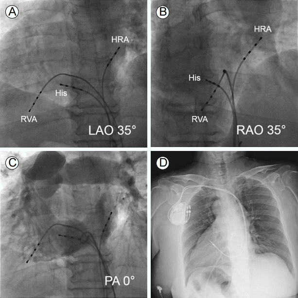
Radiographic findings. (A-C) Three quadripolar electrode catheters were placed at HRA, His, and RVA. (A) The LAO 35° view of the dextrocardia was a mirror image of the RAO view of the normal heart. (B) The RAO 35° view of the dextrocardia was similar to the LAO view of the normal heart. (C) The anatomical locations of the electrode catheters within cardiac chambers were visualized by right ventriculography in the PA 0° view. (D) An anteroposterior chest X-ray showed that the right ventricular and atrial leads were well positioned at the upper interventricular septum and right atrial appendage, respectively. HRA, high right atrium; His, His bundle area, RVA, right ventricular apex; LAO, left anterior oblique; RAO, right anterior oblique; PA, posteroanterior.
고 찰
오른심장증은 크게 내장의 위치가 정상인 경우(situs solitus)와 내장좌우바뀜증을 동반하는 경우로 나누어 볼 수 있다[1,2]. 내장의 위치가 정상인 환자에서 오른심장증이 나타날 경우 거의 대부분 선천성 심장 또는 혈관기형을 동반하며, 내장좌우바뀜증이 있는 환자에서 오른심장증이 나타날 경우 30% 미만에서만 심장 또는 혈관기형을 동반하는 것으로 알려졌다[2,8]. 오른심장증은 후천적으로 발생할 수도 있는데 폐절제술 이후에 오른심장증이 발생한 사례들이 보고된 바 있다[9,10]. 저자들의 사례처럼 심장내 기형이 동반되지 않은 오른심장증의 경우 성인까지 생존할 수 있지만, 방실전도차단이나 동기능부전 등의 서맥성 부정맥이 발생할 수 있다. 선천성 오른심장증 환자에서 영구형 인공심장박동기 거치술을 시행한 몇몇 사례가 보고되었는데 혈관과 심장내 중요 해부학적 구조물들의 위치가 정상과는 다르므로 시술에 기술적인 어려움이 따르는 것으로 알려졌다[3-7]. 특히 내장의 위치가 정상인 경우에는 교정 수술을 필요로 하는 선천성 기형인 대혈관전위(transposition of great arteries)가 동반된 경우가 많아 심장박동 조율용 유도전극을 삽입하기 어려울 수 있다[6,7]. 내장좌우바뀜증이 동반된 경우에 일반적인 환자들처럼 왼쪽 겨드랑이정맥이나 빗장뼈밑정맥(subclavian vein)을 통해 유도전극을 삽입할 경우 비정상적인 각도로 오른심실로 진입하게 되어 원하는 위치에 유도전극을 위치시키기 어려울 수 있다. 오른쪽에 위치한 겨드랑이정맥과 빗장뼈밑정맥이 해부학적으로는 왼쪽 겨드랑이정맥 및 빗장뼈밑정맥과 동일한 구조를 가지고 오른심장으로 연결되므로 오른쪽 혈관을 통해 심장박동 조율용 유도전극을 삽입하는 것이 기술적으로 타당하다. 실제로 후천적으로 발생한 오른심장증을 제외한 대부분의 선천적 오른심장증 사례들은 오른쪽 겨드랑이정맥이나 빗장뼈밑정맥을 통해 유도전극을 삽입한 것으로 보고하였다[4-7]. 오른심장증에 따른 해부학적인 장애를 극복하기 위해 영구형 인공심장박동기 거치술을 시행하기 전에 중요 혈관과 심장의 해부학적 구조를 파악하고 시술 절차를 시뮬레이션(simulation)하는 것이 시술 시간과 합병증 줄이는 데 도움이 될 수 있다. 저자들의 사례에서는 영구형 인공심장박동기 거치술을 시행하기 전에 전기 생리 검사에 사용되는 전극도자를 심장 내에 위치시킨 후 우전사위, 좌전사위, 후전위에서 방사선 투시촬영을 시행하고 오른심장조영술을 통해 심장내 중요 해부학적 구조물들의 위치를 상세하게 파악하였으며, 얻어진 방사선 영상을 길잡이로 원하는 위치에 심장박동 조율용 유도전극을 성공적으로 위치시킬 수 있었다.
오른심장증이 있는 환자를 대상으로 영구형 인공심장박동기 거치술을 시행한 증례는 흔하지 않으며 심장내 해부학적 구조물들의 위치 변화와 동반된 심장기형들로 인해 시술에 어려움이 따른다. 저자들은 완전 내장좌우바뀜증 환자를 대상으로 영구형 인공심장박동기 거치술을 시행하였기에 이를 문헌고찰과 함께 보고하는 바이다.