뼈 전이를 동반한 진행성 위암 환자에서의 Flare Phenomenon
The Flare Phenomenon in a Patient with Advanced Gastric Cancer with Bone Metastases
Article information
Trans Abstract
Flare phenomenon refers to increased radiotracer uptake in bones despite clinical findings showing a positive response to treatment. Flare phenomena are most often observed in patients with breast or prostate cancer. Here, we present a case of bone flare in a 54-year-old male who had advanced gastric cancer with bone metastases. After three cycles of chemotherapy, a bone scan showed increased intensity, but the patient’s bone pain was alleviated and abdominal computed tomography revealed a decrease in the size of the primary mass and metastatic lymph nodes. We therefore continued chemotherapy using the same regimen, and a follow-up bone scan revealed decreased intensity. A flare phenomenon after treatment is rare in cases of gastric cancer with bone metastasis. Although flare phenomena are not common, they should be considered in patients with gastric cancer when the clinical results are inconsistent with bone-scan findings.
INTRODUCTION
Bone metastasis is a devastating morbidity in patients with advanced cancer. In addition to causing pain, the condition may lead to immobility, pathological bone fracture, and cord compression.
Although bone metastases are more common in prostate, breast, and lung cancers, they are not rare in patients with gastric cancer. A recent Korean study found that the incidence of bone metastases from metastatic or recurrent gastric cancer was approximately 11% [1].
Bone scanning using Technetium-99m hydroxydiphosphonate (HDP) is a nuclear imaging technique commonly used to detect bone metastases [2]. The flare phenomenon is defined as bone-scan and serum alkaline phosphate (ALP) findings that show disease progression after treatment despite indications of a good therapeutic response in terms of clinical symptoms or decreased tumor size on computed tomography (CT) scans. The bone-scan flare phenomenon was first described in 1972 by Greenberg et al. [3]. Although it is difficult to distinguish flare phenomena from disease progression during treatment, the distinction is critical as misinterpretation may lead to inappropriate decisions regarding the course of treatment. Here, we describe a case of a flare phenomenon in a 54-year-old male who was receiving chemotherapy for gastric cancer with bone metastasis.
CASE REPORT
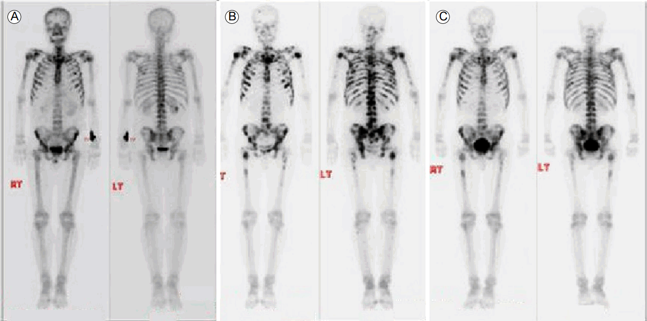
A 54-year-old male patient was admitted because of tarry stool. Esophagogastroduodenoscopy showed bleeding from a gastric mass, and serial biopsy revealed poorly differentiated adenocarcinoma. A complete blood count showed a hemoglobin of 7.7 g/dL (normal range: 13-17 g/dL), and a laboratory study showed a serum level ALP of 133 IU/L (normal range: 40-129 IU/L). Abdominal CT scan revealed a bulky mass and accompanying extraserosal extension at the gastric body and antrum. The lymph nodes were markedly enlarged at the esophagogastric junction, and in the left gastric, porta hepatis, peripancreatic celiac, superior mesenteric artery, and para-aortic regions, and peritoneal thickening with ascites was evident (Fig. 1A). Positron emission tomography (PET) showed multiple hypermetabolic lymph nodes and hypermetabolic peritoneal thickening in the same lesion was observed on CT. Furthermore, PET revealed extensive hypermetabolic lesions involving the entire spine, sternum, both sides of the rib cage, the clavicle, the scapulae, the humerus and femur, and the pelvic bone. These lesions were identified as multiple bone metastases (Fig. 2). The bone scan showed increased heterogeneous uptake (Fig. 3A).

Enhanced abdomen computed tomography (CT) axial images at baseline (A), after the initial three cycles (B), and additional three cycles (C) of chemotherapy. Baseline CT revealed a bulky mass with extraserosal extension at the gastric body and antrum. After chemotherapy, CT images showed a continuous decrease in the bulky mass.

Positron emission tomography with concurrent computed tomography revealed hypermetabolic lesions involving the entire spine, sternum, both sides of the rib cage, clavicle, scapulae, humerus and femur, and pelvic bone.

Technetium-99m hydroxydiphosphonate (99mTc-HDP) bone scans at baseline (A), after the initial three cycles (B), and additional three cycles (C) of chemotherapy. The baseline bone scan showed increased 99mTc-HDP uptake throughout the skeleton. We observed an increase in the intensity, size and number of metastases after the initiation of chemotherapy. Following treatment with the additional cycles of chemotherapy, the bone scan showed reductions in the intensity and extent of uptake in the multiple metastases.
Chemotherapy was planned to treat the cancer. The folinic acid (leucovorin) + fluorouracil (5-FU) + oxaliplatin (eloxatin) (FOLFOX) regimen was administered at 2-week intervals (oxaliplatin, 100 mg/m2 as a 2-h infusion; leucovorin, 400 mg/m2 as a 2-h infusion; and a 5-fluorouracil bolus, 400 mg/m2, followed by a 5-fluorouracil continuous infusion, 1200 mg/m2 in 22 h). After three cycles of chemotherapy, abdominal CT, bone scan, and laboratory tests were performed. Serum ALP was elevated continuously up to 2,038 IU/L, and the bone scan revealed increased intensity of the multiple bone metastases during the same period (Fig. 3B). However, abdominal CT revealed a marked reduction in the primary mass on the gastric body and antrum and nearby lymph nodes, and improvement in the peritoneal cancer (Fig. 1B). We reduced the dose of narcotic analgesics because the patient reported that the bone pain was alleviated throughout his body. Although the bone-scan and serum ALP findings suggested worsening of the bone metastases, we concluded that these findings were the result of the flare phenomenon because the patient’s symptoms and the CT findings indicated that his condition had improved. Accordingly, we continued treatment without changes to the chemotherapy regimen.
After three additional cycles of chemotherapy, abdominal CT revealed further reductions in the gastric mass and lymph node metastases (Fig. 1C). A bone scan showed reductions in the intensity and extent of uptake in the multiple bone metastases (Fig. 3C), and the serum ALP level decreased to 510 IU/L (i.e., 1/4 of the previous value), which indicated improvement in the bone metastases. Consequently, the therapy was continued, and the patient received six cycles of FOLFOX without further complications. However, the patient died from hospital-acquired pneumonia when he was admitted for the seventh cycle of FOLFOX.
DISCUSSION
We describe a rare case of flare phenomenon in a patient with advanced gastric cancer with bone metastases. Bone metastases frequently occur in patients with advanced primary cancers of the breast, prostate, lung, and kidney; however, the condition is less common in cases of gastric cancer, and the prognosis is poor. The peritoneum, liver, and lymph nodes are common metastatic sites of gastric cancer. Nakanishi et al. [4] found that bone metastases occurred in 1 to 11% of patients with gastric cancer [5]. Ahn et al. [5] reported that the prevalence of bone metastases was about 0.9% in patients with gastric cancer, and that the most common metastatic sites were the thoracic and lumbar vertebrae [4,5].
Bone scans are generally performed to detect bone metastases and evaluate treatment response. Bone scans show an increase in radiotracer uptake when bone is actively formed. Serum ALP, which is related to bone growth, is present in several cell membranes (including osteoblastic cells) and is released into the blood during bone formation. Elevated serum ALP levels at the time of gastric cancer diagnosis is an indicator of bone metastasis, and the therapeutic response can be monitored by measuring serum ALP levels after treatment. Increased tracer uptake is a marker of osteoblastic activity. Cancer treatment shrinks tumors in the bone leading to osteoblastic changes. Thus, a flare phenomenon is thought to result from transient increases in bone-specific radiotracer uptake (such as of HDP) at the metastatic lesion, owing to osteoblastic healing [6].
The flare phenomenon was first described by Greenberg et al. [3] in 1972, and similar cases have been reported in Europe and the United States [3,7,8]. The flare phenomenon occurs most frequently in patients with breast or prostate cancer, early in the treatment of bone metastases. Several case studies have reported that bone flare appeared after treatment in patients with lung cancer; however, the frequency of flare phenomenon in lung cancer has not been thoroughly investigated [9].
Given that the presence of a flare on a bone scan and elevated serum ALP levels suggest a transient worsening of the patient’s condition, it is critical that the flare phenomenon is distinguished from true disease progression because misdiagnosis can lead to early discontinuation of therapy or inappropriate changes in the treatment regimen. Bone scans and serum ALP levels are not specific indices of bone metastases; thus, it is necessary to obtain additional CT and magnetic resonance imaging scans and integrate all the clinical data including imaging, patient symptoms and physical examination results. Our patient’s back pain was relieved and the status of his primary cancer improved on abdominal CT, and the case was judged to reflect a flare phenomenon.
In conclusion, the flare phenomenon is rare in the early treatment of gastric cancer with bone metastasis. Although bone-scan and serum ALP findings are useful for detecting bone metastases and evaluating therapeutic response, these findings may, paradoxically, indicate disease progression in patients with a good therapeutic response, indicating a situation known as the bone flare phenomenon. Although it is difficult to distinguish between a bone flare and true progression of disease activity, particularly in the short term, this distinction is critical because it may influence the overall treatment course.