Korean J Med > Volume 93(4); 2018 > Article
아칼라지아의 새로운 분류법: Chicago's Classification

요약

고해상도 식도내압 검사는 우리에게 식도운동에 대한 보다 세밀하고 다양한 정보를 제공할 수 있으며 식도의 병태생리에 대한 이해를 높이는데 중요한 역할을 하고 있다. 식도운동 질환의 경우 역류성 식도 질환이나 다른 심장 질환과 감별이 쉽지 않아 진단이 늦어지는 경우가 많았으나 새로운 검사장비와 분류법으로 인하여 질환의 조기 진단이 가능해졌으며 그동안 우리가 알지 못했던 식도의 비정상 상태를 파악할 수 있게 되어 식도운동 질환 환자의 삶의 질을 높일 수 있으리라 기대된다.

증 례

27세 남자가 1년 전부터 발생한 역류 증상, 가슴답답함 및 체중감소로 내원하였다. 6개월 전 개인의원에서 내시경 검사를 시행하여 역류성식도염 외 특이 소견이 없어 proton pump inhibitor (PPI)를 포함한 약물을 3개월간 복용하였으나 증상이 더욱 심해져 최근에는 물도 삼키기 힘들어졌다고 한다.

진단의 일차 단계

본원에서 시행한 내시경에서 위식도접합 부위에 미란 등은 관찰되지 않았고 위식도접합부(esopahgogastric junction, EGJ) 통과 시 약간의 저항감만 느껴졌다. PPI를 장기간 복용함에도 불구하고 증상이 오히려 악화되었으며, 최근에는 삼킴곤란 증상까지 발생하여 식도운동 질환이 의심되어 고해상도 식도내압 검사를 시행하였다. 고해상도 식도내압 검사상 하부식도 괄약근의 이완시 압력인 integrated relaxation pressure (IRP) 값이 24.7 mmHg로 상승되어 있었으며 식도연동운동은 소실되었고 panesophageal pressurization 소견을 보였다(Fig. 1).

최종 진단과 치료 경과

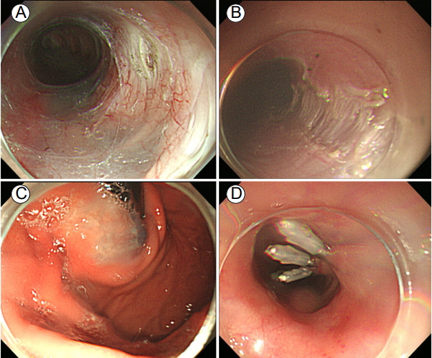
고해상도 식도내압 검사상 type II achalasia로 진단되었고 환자의 Eckardt 증상 점수는 11점이었다. 치료 계획으로 풍선확장술과 경구 내시경 근절개술(peroral endoscopic myotomy, POEM)을 고려하였으며 환자와 상의하여 경구 내시경 근절개술을 시행하기로 하였다. 경구 내시경 근절개술은 전신마취 하에 수술실에서 진행되었고 위식도접합부 하부 2 cm를 포함하여 총 14 cm의 길이로 근절개를 시행하였다(Fig. 2). 시술 후 환자의 Eckardt 증상 점수는 11점에서 1점으로 감소하였고 고해상도 식도내압 검사 소견도 IRP 값이 24.7 mmHg에서 7.4 mmHg로 감소하였다(Fig. 3). 시술 후 1년 경과 시까지 증상의 재발이 보이지 않고 잘 유지되고 있다.

고 찰

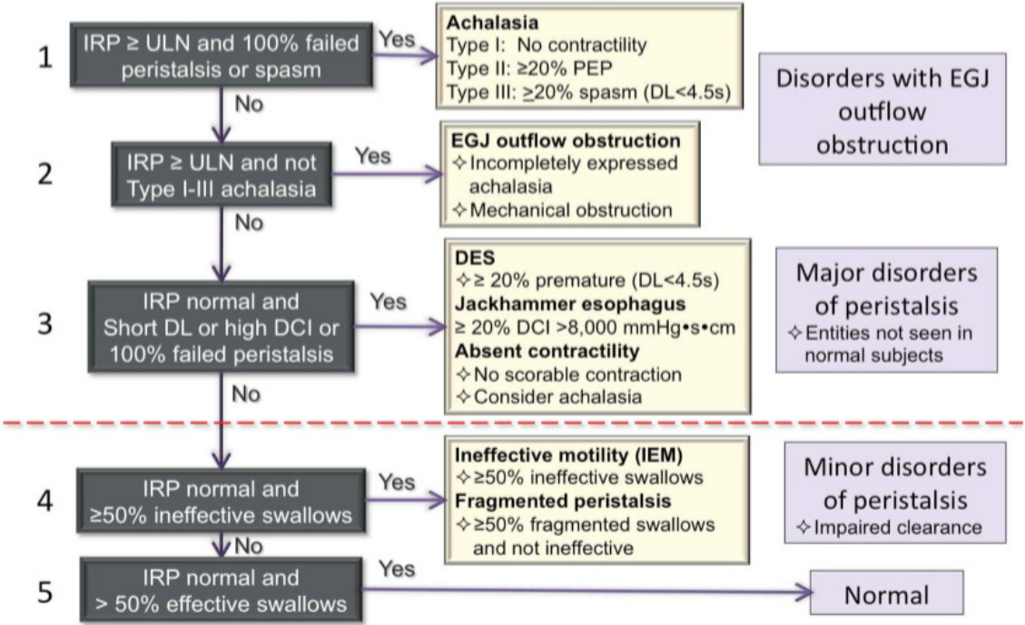
고해상도 식도내압 검사는 기존의 식도내압 검사에 비하여 압력 센서가 치밀하게 분포하여 식도운동 변화를 보다 세밀하게 측정할 수 있다. 이 기기의 도입으로 식도운동 질환의 진단에 있어 획기적인 발전이 이루어졌을 뿐 아니라 식도운동 질환의 정의와 분류마저 새로이 정립되었다. 고해상도 식도내압 검사 결과를 기반으로 식도운동 질환의 새로운 분류법이 만들어지게 되었는데 처음 버전은 2008년 샌디에고에서 열렸던 International HRM Working Group 회의를 통하여 제시되었다[1]. 이후 몇 번의 수정을 거쳐 2014년 Chicago에서 세계 각국의 소화기운동학회 관련 연구자들이 모여 회의한 결과 지금 현재 사용되고 있는 Chicago classification v3.0이 만들어지게 되었다[2]. 고해상도 식도내압 검사에서는 압력 수치 이외에 다양한 지표들이 측정 가능한데(Table 1) [2], 이러한 지표들은 크게 수축 강도, 수축 패턴, 삼킴 시 30 mmHg 등압 윤곽선 패턴에 따라 여러 가지의 비정상 운동 패턴으로 분류된다(Table 2) [2]. Chicago classification은 이러한 여러 가지 지표와 운동 패턴을 계층적으로 분석하여 다양한 형태의 식도운동 질환을 진단할 수 있도록 도와준다(Fig. 4) [3]. 아칼라지아의 경우 Chicago classification에 따라 1형, 2형, 3형으로 분류할 수 있으며 식도체부의 연동운동은 정상이지만 하부식도괄약근의 이완에 장애가 있는 위식도접합부 폐쇄(EGJ outflow obstruction, EGJOO)라는 새로운 카테고리의 질환도 진단할 수 있게 되었다. 업데이트된 Chicago classification을 적용할 경우 기존의 분류법에 비하여 진단이 불명확한 경우와 주요 운동 질환(major motor disorder)의 빈도가 줄어든 대신 위식도접합부 폐쇄나 사소한 운동 질환(minor motor disorder)의 빈도가 늘어난 것으로 보고되고 있다[3].
고해상도 식도내압 검사 결과를 기반으로 한 Chicago classification을 사용하였을 경우 아칼라지아를 비롯한 여러 식도운동 질환을 진단하는데 있어 기존의 식도내압 검사의 진단 기준을 사용하였을 때에 비하여 평가자 간 진단 일치도도 높고 배우기도 쉬우며 숙련자나 비숙련자 모두 진단 정확도가 높아지는 것으로 보고되고 있다[4-6]. 또한 고해상도 식도내압 검사를 기반으로 한 Chicago classification이 점차 널리 사용되고 있으며 식도운동 질환의 진단에 최적 표준으로 간주되고 있다.

REFERENCES

1. Pandolfino JE, Fox MR, Bredenoord AJ, Kahrilas PJ. High-resolution manometry in clinical practice: utilizing pressure topography to classify oesophageal motility abnormalities. Neurogastroenterol Motil 2009;21:796–806.
crossref pmid pmc

2. Kahrilas PJ, Bredenoord AJ, Fox M, et al. The Chicago classification of esophageal motility disorders, v3.0. Neurogastroenterol Motil 2015;27:160–174.
crossref pmid

3. Laing P, Bress AP, Fang J, Peterson K, Adler DG, Gawron AJ. Trends in diagnoses after implementation of the Chicago classification for esophageal motility disorders (v3.0) for high-resolution manometry studies. Dis Esophagus 2017;30:1–6.
crossref

4. Fox MR, Pandolfino JE, Sweis R, et al. Inter-observer agreement for diagnostic classification of esophageal motility disorders defined in high-resolution manometry. Dis Esophagus 2015;28:711–719.
crossref pmid

5. Carlson DA, Ravi K, Kahrilas PJ, et al. Diagnosis of esophageal motility disorders: esophageal pressure topography vs. conventional line tracing. Am J Gastroenterol 2015;110:967–977.
crossref pmid pmc

6. Soudagar AS, Sayuk GS, Gyawali CP. Learners favour high resolution oesophageal manometry with better diagnostic accuracy over conventional line tracings. Gut 2012;61:798–803.
crossref pmid

High resolution manometry before POEM procedure. High resolution esophageal pressure topography shows increased IRP (24.7 mmHg) and panesophageal pressurization. POEM, peoral endoscopic myotomy; IRP, integrated relaxation pressure.
/upload/thumbnails/kjm-93-4-360f1.jpg
Figure 1.
POEM procedure. (A) Submucosal tunneling. (B) Myotomy. (C) Retroflexion view of cardia. (D) Incision site closure with hemoclipp. POEM, peoral endoscopic myotomy.
/upload/thumbnails/kjm-93-4-360f2.jpg
Figure 2.
High resolution manometry after POEM procedure. High resolution esophageal pressure topography shows decreased IRP (7.4 mmHg) after POEM. POEM, peoral endoscopic myotomy; IRP, integrated relaxation pressure.
/upload/thumbnails/kjm-93-4-360f3.jpg
Figure 3.
Hierarchial analysis of Chicago classification v3.0 [3]. IRP, integrated relaxation pressure; ULN, upper limit of normal; PEP, panesophageal pressurization; DL, distal latency; EGJ, esophagogastic juction; DCI, distal contractile integral; DES, distal esophageal spasm.
/upload/thumbnails/kjm-93-4-360f4.jpg
Figure 4.
Table 1.
Pressure topography metric utilized in the Chicago classification v3.0 unless otherwise specified, pressures are referenced to atmospheric [2]
Pressure topography metrics
CDP (time, position)a Inflection point along the 30 mmHg isobaric contour (or pressure greater than intrabolus pressure in instances of compartmentalized pressurization) at which propagation velocity slows, demarcating peristalsis from ampullary emptying. The CDP must be localized within 3 cm of the proximal margin of the LES
DCI (mmHg·s·cm)b Amplitude × duration × length (mmHg·s·cm) of the distal esophageal contraction exceeding 20 mmHg from the transition zone to the proximal margin of the LES (Clouse, 2nd and 3rd contractile segments)c
DL (s)a Interval between UES relaxation and the CDP
IRP (mmHg)b Mean of the 4 s of maximal deglutitive relaxation in the 10-s window beginning at UES relaxation. Contributing times can be contiguous or non-contiguous (e.g., interrupted by diaphragmatic contraction). Referenced to gastric pressure.

CDP, contractile deceleration point; LES, low esophageal sphincter; DCI, distal contractile integral; DL, distal latency; IRP, integrated relaxation pressure; UES, upper esophageal sphincter.

a Normal values are independent of the manometric hardware utilized.

b Normal values are dependent on the speciftc manometric hardware utilized.

c In instances of suspected hypercontractility involving the LES, or even restricted to the LES, the DCI box should include the LES (Clouse, 4th contractile segment) as well.

Table 2.
Characterization of esophageal contractility: contraction pattern is not scored for ineffective swallows (DCI < 450 mmHg) [2]
Contraction vigor
 Failed DCI < 100 mmHg·s·cm
 Weak DCI > 100 mmHg·s·cm, but < 450 mmHg·s·cm
 Ineffective Failed or weak
 Normal DCI > 450 mmHg·s·cm but < 8,000 mmHg·s·cm
 Hypercontractile DCI ≥ 8,000 mmHg·s·cm
Contraction pattern
 Premature DL < 4.5 s
 Fragmented Large break (> 5 cm length) in the 20-mmHg isobaric contour with DCI > 450 mmHg·s·cm
 Intact Not achieving the above diagnostic criteria
Intrabolus pressure pattern (30 mmHg isobaric contour referenced to atmospheric)
Panesophageal pressurization Uniform pressurization of > 30 mmHg extending from the UES to the EGJ
 Compartmentalized Pressurization of > 30 mmHg extending from the contractile
 Esophageal pressurization Front to the EGJ
 EGJ pressurization Pressurization restricted to zone between the LES and CD in conjunction with LES-CD separation
 Normal No bolus pressurization > 30 mmHg

DCI, distal contractile integral; DL, distal latency; UES, upper esophageal sphincter; EGJ, esophagogastic juction; LES, low esophageal sphincter; CD, crural diaphragm.

TOOLS
METRICS Graph View
  • 0 Crossref
  •  0 Scopus
  • 9,811 View
  • 372 Download

Editorial Office
101-2501, Lotte Castle President, 109 Mapo-daero, Mapo-gu, Seoul 04146, Korea
Tel: +82-2-2271-6791    Fax: +82-2-790-0993    E-mail: kaim@kams.or.kr                

Copyright © 2025 by The Korean Association of Internal Medicine.

Developed in M2PI

Close layer
prev next