

INTRODUCTION
Left atrial (LA) dissection is a rare but potentially serious complication of cardiac surgery and is most commonly reported following mitral valve (MV) procedures [1-3]. We report the case of a 76-year-old woman who developed LA dissection after endoatriectomy for severe “eggshell” calcification of the LA wall, performed concurrently with bioprosthetic MV replacement (MVR) and a maze procedure. To our knowledge, this is the first reported case of this complication in Korea. Notably, whereas most previously reported cases required surgical repair, the LA dissection in our patient re-solved spontaneously with conservative management. This case underscores the importance of recognizing LA dissection as a potential complication of endoatriectomy and suggests that careful nonoperative management can be a safe alternative in hemodynamically stable patients.
CASE REPORT
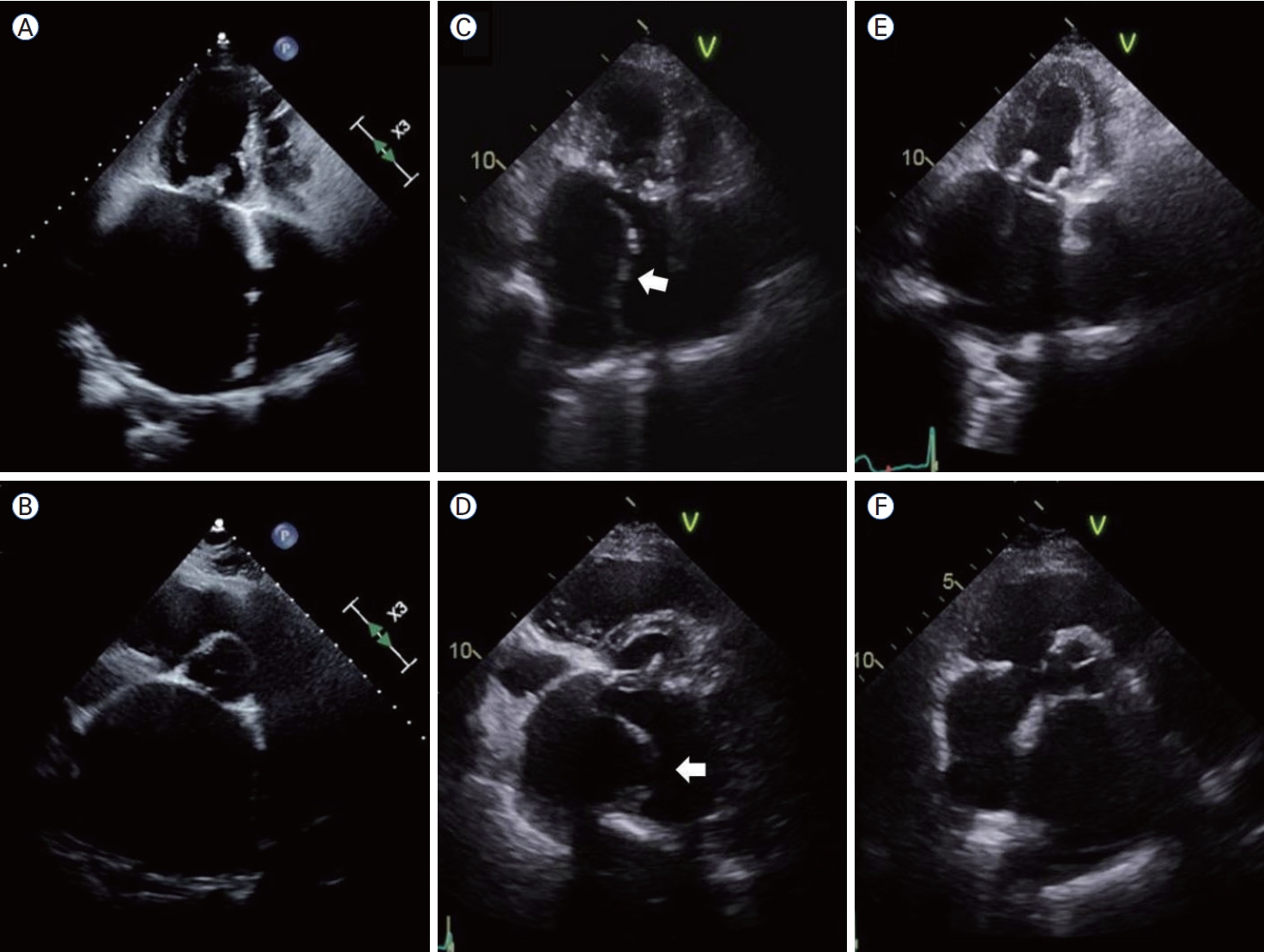
A 76-year-old woman with hypertension and rheumatic mitral stenosis (MS) presented with exertional dyspnea and palpitations. Chest radiography demonstrated marked cardiomegaly, pulmonary congestion, and curvilinear calcification along the LA wall (Fig. 1A, B). Electrocardiography showed atrial fibrillation (AF) with a rapid ventricular response. Transthoracic echocardiography revealed severe rheumatic MS with mild mitral regurgitation (MV area of 0.92 cm2, mean transmitral pressure gradient of 17.8 mmHg), marked enlargement of the LA (LA volume index of 205.4 mL/m2, LA diameter of 85 mm), and moderate tricuspid regurgitation with tricuspid annular dilatation (Fig. 2A, B). Cardiac computed tomography (CT) showed circumferential eggshell calcification of the LA (Figs. 1C, 3A, B).
The patient underwent bioprosthetic mitral valve replacement for MS, endoatriectomy, maze procedure with LA appendage obliteration, and tricuspid annuloplasty. The immediate postoperative clinical course was uneventful, and her symptoms improved significantly.
Ten days later, follow-up echocardiography revealed a sheet-like lesion separating the true LA lumen from a large cystic false lumen extending along the atrial wall, consistent with LA dissection (Fig. 2C, D; Supplementary Video 1). The false lumen did not cause hemodynamic compromise, including obstruction of blood flow through the prosthetic MV. Cardiac CT demonstrated a thrombus-filled dissection involving the inferior and septal areas of the LA wall without pulmonary venous involvement (Fig. 3C, D). Because the patient remained clinically stable without any symptoms or signs of heart failure, and echocardiography revealed a well-functioning prosthetic MV without blood flow obstruction, conservative management with close clinical monitoring and imaging studies was pursued. After 2 weeks of medical management, repeat CT showed no significant change. Warfarin therapy for AF was continued during this period. The patient was discharged on warfarin (target international normalized ratio, 2.0-3.0) and bisoprolol for heart rate control. Three months later, anticoagulation therapy was switched from warfarin to rivaroxaban (20 mg daily). Over 6 months, serial echocardiography (Fig. 2E, F; Supplementary Video 2) and cardiac CT (Fig. 3E, F) documented gradual regression and eventual complete resolution of the LA dissection.
DISCUSSION
LA dissection is a rare postoperative complication in which a false or true lumen forms between the endocardial and myocardial layers, most commonly after MV surgery [1-3]. It is generally associated with iatrogenic injury or disruption of the LA endocardium during procedures involving aggressive decalcification or atrial wall manipulation. It may lead to hemodynamic compromise when the mitral inflow or pulmonary venous return is obstructed. In this patient, circumferential eggshell calcification likely made the atrial wall more fragile, and endoatriectomy further increased susceptibility to wall separation [4-6]. In addition, the mechanical stress associated with MVR and postoperative hemodynamic changes cannot be excluded as contributing factors, even in the absence of prosthetic valve dysfunction or paravalvular leak.
In patients with long-standing rheumatic mitral valve disease, extensive LA calcification reflects advanced chronic atrial remodeling and has been associated with limited efficacy of the maze procedure. Nevertheless, endoatriectomy may be considered in selected patients to facilitate surgical exposure during MV surgery, reduce the thrombogenic atrial surface, and allow more effective transmural energy delivery during the maze procedure [7]. In this context, the development of LA dissection in our patient may reflect the underlying atrial pathology and complexity of the surgical procedure.
Diagnosis of LA dissection relies on high-resolution cardiac imaging. Echocardiography is useful for identifying dissection and evaluating hemodynamic consequences, although it may be insufficient for defining its full extent and associated complications. Transesophageal echocardiography is often preferred for delineating the dissection plane and assessing involvement of the prosthetic valve or pulmonary venous inflow [3,8]. Color Doppler imaging further confirmed the absence of turbulent or high-velocity flow, suggesting significant communication between the true and false lumens. In the present case, cardiac CT served as an alternative, providing clear delineation of the false lumen and thrombus. Multimodal cardiovascular imaging is therefore essential for evaluating the presence, anatomical extent, and hemodynamic impact of LA dissection and guiding therapeutic decisions.
The management of LA dissection is not standardized and depends on individual patient-specific factors, including symptoms, hemodynamic status, anatomical extent, and associated valvular or structural complications [3]. Surgical correction is indicated in cases of hemodynamic instability, flow obstruction, or prosthetic valve dysfunction. However, stable patients without obstruction may be managed conservatively through blood pressure control, rhythm monitoring, appropriate anticoagulation, and serial imaging surveillance. Conservative therapy may be a viable alternative to surgery in stable patients, particularly in elderly individuals with high operative risk or complex surgical anatomy. Nonetheless, a multidisciplinary team involving cardiologists, cardiac surgeons, and imaging specialists is essential for individualized decision making.
In a comprehensive review by Tsukui et al [3]., most cases required surgical repair such as re-exploration, patch repair, or even valve re-replacement, especially when associated with hemodynamic deterioration, mechanical obstruction of the mitral or pulmonary venous inflow tracts, or systemic embolism. Only a few cases of spontaneous resolution without reoperation have been reported. Alshaabi et al [8]. reported an intraoperative LA dissection after mitral repair requiring immediate patch repair, whereas Bidmead et al [9]. reported a postoperative case that was successfully managed conservatively with a favorable outcome. Our patient’s course supports the latter, suggesting that nonoperative treatment may be safe in hemodynamically stable asymptomatic patients without significant obstruction or thromboembolic risk. Our experience further supports the feasibility of nonoperative management, even after complex procedures such as endoatriectomy for extensive eggshell calcification combined with MVR.
As a single case report, our findings are observational and cannot be generalized. Moreover, although our patient showed spontaneous resolution, long-term follow-up is required to monitor for potential complications, including atrial arrhythmias, thrombus formation, or late structural remodeling. The lack of histopathologic confirmation is inherent in the nonsurgical approach, and diagnosis relies solely on imaging findings. However, this case highlights the potential for spontaneous resolution of LA dissection following endoatriectomy and MVR. Awareness of this rare complication, particularly in patients with rheumatic mitral disease and extensive atrial calcification, is crucial for timely diagnosis and appropriate management.
In conclusion, we describe a rare case of LA dissection following endoatriectomy for eggshell calcification combined with MVR, which was successfully treated conservatively. Unlike most previously reported cases that necessitate surgical repair, the dissection in our patient resolved without complications. Our findings reinforce the importance of individualized assessment and the potential for nonoperative treatment in hemodynamically stable patients. With an increasing number of elderly and high-risk patients undergoing complex cardiac surgery, awareness of LA dissection and its variable clinical course is essential for optimal patient outcomes. The accumulation of further cases is essential to refine the diagnostic strategies and management algorithms for this rare but potentially serious complication.




 PDF Links
 PubReader
 ePub Link
 XML Download
 Full text via DOI
 Download Citation
 Print
Print