호흡 곤란과 실신으로 내원한 환자에서 정맥혈전색전증의 진단

A Diagnostic Roadmap for Venous Thromboembolism: Pulmonary Embolism and Deep Vein Thrombosis

Article information

Korean J Med. 2026;101(5):255-266
Publication date (electronic) : 2026 October 1
doi : https://doi.org/10.3904/kjm.2026.101.5.255
Division of Cardiovascular, Department of Internal Medicine, Kyung Hee University Hospital, Kyung Hee University School of Medicine, Seoul, Korea
우종신
경희대학교 의과대학 경희대학교병원 심장내과
Correspondence to: Jong Shin Woo, M.D., Ph.D. Division of Cardiovascular, Department of Internal Medicine, Kyung Hee University Hospital, Kyung Hee University School of Medicine, 23 Kyungheedae-ro, Dongdaemun-gu, Seoul 02447, Korea Tel: +82-2-958-8170, Fax: +82-2-958-8160, E-mail: cardiodrwoo@gmail.com
Received 2026 June 22; Revised 2026 August 23; Accepted 2026 September 21.

Trans Abstract

Venous thromboembolism (VTE), encompassing pulmonary embolism (PE) and deep vein thrombosis (DVT), requires a structured diagnostic roadmap to prevent delayed diagnosis and unnecessary imaging. This review outlines a step-by-step approach from clinical suspicion to objective confirmation and early risk stratification. For suspected PE, hemodynamic stability is the initial branch point. Hemodynamically stable patients are evaluated using pretest probability tools and validated D-dimer strategies, including age-adjusted thresholds, YEARS, or clinical-probability-adjusted approaches such as PEGeD, followed by computed tomography pulmonary angiography when indicated. Pregnancy-adapted YEARS can reduce thoracic imaging in selected pregnant patients. For suspected DVT, the Wells score, D-dimer, and compression ultrasonography are applied sequentially. Once PE is confirmed, risk assessment should integrate clinical severity, right ventricular dysfunction, cardiac biomarkers, hemodynamic and respiratory compromise to guide management decisions. The final diagnosis should specify thrombus location and extent, risk category, provoking factors, and remaining diagnostic uncertainty. Standardizing this roadmap may reduce diagnostic delay and unnecessary imaging while linking diagnosis to appropriate disposition, monitoring, and follow-up.

증례: 79세 남자가 실신 후 응급실에 내원하였다. 의식 소실은 일시적이었고 내원 시 신경학적 결손은 뚜렷하지 않았으나 호흡 곤란과 전신 쇠약감을 호소하였다. 활력 징후는 혈압 156/78 mmHg, 심박수 85회/분, 호흡수 20회/분, 실내 공기 산소포화도 80%였으며, 비강 캐뉼라로 산소 5 L/min을 투여한 후 산소포화도는 96%로 유지되었다. 고혈압, 당뇨병 및 관상동맥질환의 과거력이 있었고 aspirin, amlodipine/atorvastatin, carvedilol, fimasartan, doxazosin 및 dapagliflozin/metformin 을 복용 중이었다. 심전도에서 ST분절 상승은 없었다. 고감도 트로포닌 I (high-sensitivity troponin I, hs-troponin I)은 28.6 pg/mL로 99백분위 상한 기준치(17.5 pg/mL)를 초과하였고, D-dimer는 >20 μg/mL (참고치 < 0.5 μg/mL), N-말단 B형 나트륨이뇨펩타이드(N-terminal pro-B-type natriuretic peptide, NT-proBNP)는 336 pg/mL, 젖산은 2.15 mmol/L, 추정 사구체여과율은 50.12 mL/min/1.73 m2였다. 폐동맥 컴퓨터단층혈관조영술에서 양측 주폐동맥과 양측 폐의 다발성 엽, 분절 폐동맥 가지를 침범하는 급성 폐색전증이 확인되었다. 심초음파에서는 우심실/좌심실 비 0.9, 삼첨판륜 수축기 이동거리(tricuspid annular plane systolic excursion, TAPSE) 15.9 mm, 우심실 분획면적 변화(right ventricular fractional area change, RVFAC) 18%, 추정 폐동맥 수축기압 43.8 mmHg였다. 하지 압박초음파에서는 양측 슬와정맥의 혈전이 확인되었다.

서 론

정맥혈전색전증(venous thromboembolism, VTE)은 심부정맥혈전증(deep vein thrombosis, DVT)과 폐색전증(pulmonary embolism, PE)을 포함하는 하나의 질환 스펙트럼이다. 하지 심부정맥의 혈전은 통증, 부종, 압통과 같은 국소 증상을 유발하고, 일부는 폐동맥으로 이동하여 호흡 곤란, 흉통, 실신, 저산소증, 쇼크 또는 돌연사로 발현한다[1-4]. 심부정맥혈전증과 폐색전증은 병태생리적으로 연결되어 있지만 실제 진단의 핵심 검사는 각각 압박초음파(compression ultraso-nography, CUS)와 폐동맥 컴퓨터단층혈관조영술(computed tomography pulmonary angiography, CTPA)로 다르므로 두 진단 경로를 명확히 구분할 필요가 있다[1-4].

정맥혈전색전증의 진단이 어려운 이유는 임상 양상이 비특이적이기 때문이다. 폐색전증은 전형적인 호흡 곤란이나 흉통 없이 실신, 빈맥, 원인 불명의 저산소증, 심부전 악화 또는 쇠약감으로 나타날 수 있다[2,4]. 하지 부종은 봉와직염, 만성 정맥부전, 림프부종, 근골격계 손상 또는 Baker 낭종 파열과의 감별이 필요하다[1,3,5]. 진단이 지연되면 치명적인 폐색전증을 놓칠 수 있는 반면 임상 가능성이 낮은 환자에게 무분별하게 영상 검사를 시행하면 방사선 및 조영제 노출, 우연히 발견되는 소견, 아분절 혈전의 과잉 진단과 불필요한 항응고 치료를 초래할 수 있다[1,2,4]. 따라서 의심 환자의 진단을 놓치지 않으면서 정맥혈전색전증 가능성이 낮은 환자에게 영상 검사가 무분별하게 사용되지 않도록 하는 것이 중요하다[1-4].

본고에서는 정맥혈전색전증의 증상과 위험 인자, 검사 전 임상 가능성 평가 방법, D-dimer와 영상 검사의 활용 그리고 입원의 필요성과 모니터링 수준을 결정하는 초기 위험도 분류를 포함한 진단 로드맵을 제시하고자 한다. 구조화된 진단 로드맵은 위험도를 빠르게 평가할 수 있는 틀을 제공함으로써 진단 지연을 줄이고 임상 가능성에 따라 적절한 영상 검사를 선택하도록 하여 불필요한 검사를 줄이며 확진 후 위험 수준을 체계적으로 평가하여 입원, 모니터링 및 초기 치료 전략을 효율적으로 결정하는 데 도움을 준다[1-4,6-12].

문헌 고찰 방법

본고는 체계적 문헌 고찰이 아닌 임상 적용을 목적으로 한 서술적 문헌 고찰이다. PubMed/MEDLINE과 주요 학회 및 기관의 공식 지침을 중심으로 2013년 1월부터 2026년 7월까지 발표된 문헌을 검토하였고 Wells 점수, 폐색전증 배제 기준(Pulmonary Embolism Rule-out Criteria, PERC), 폐색전증 중증도 지수(Pulmonary Embolism Severity Index, PESI)와 같이 현재 진료의 근거가 된 고전적 도출과 검증 연구는 기간 제한 없이 포함하였다. 주요 검색어는 “venous thromboembolism,” “pulmonary embolism,” “deep vein thrombosis,” “pretest probability,” “D-dimer,” “CT pulmonary angiography,” “compression ultrasonography,” “YEARS,” “PEGeD,” “pregnancy,” “PESI,” “sPESI,” “Hestia” 및 “follow-up”이었다.

근거 선택에서는 진료지침과 전문가 합의문, 전향적 진단관리 연구, 다기관 코호트, 무작위 임상시험 및 고품질 메타분석을 우선하였다. 단일 증례 보고, 소아만을 대상으로 한 연구, 상지 심부정맥혈전증 또는 특수 시술 관련 혈전만을 다룬 연구는 일반적인 진단 로드맵과 직접 관련이 없는 경우 제외하였다. 서로 다른 진단 전략이 존재하는 영역에서는 특정 절단값을 임의로 혼합하기보다 하나의 전향적으로 검증된 알고리즘을 일관되게 적용하는 원칙을 채택하였다.

본 론

진단 로드맵의 출발점: 임상 가능성 평가

정맥혈전색전증의 진단은 검사에서 시작하지 않는다. 최근 수술 또는 외상, 3일 이상의 부동, 활동성 암, 과거 정맥혈전색전증, 임신 또는 산욕기, 에스트로겐 사용, 중심정맥관, 비만, 항인지질증후군 및 만성 염증성 질환과 증상을 함께 고려하여 검사 전 임상 가능성을 추정한다(Table 1) [1-3,13,14]. 예방적 항응고를 시행하였거나 수술 후 퇴원한 상태라고 해서 정맥혈전색전증을 완전히 배제할 수 있는 것은 아니다[3,13,14].
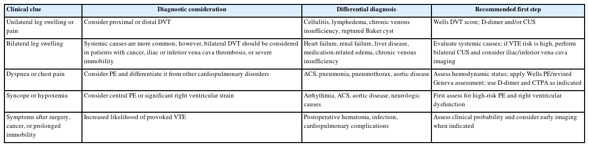
Clinical clues prompting suspicion of venous thromboembolism

심부정맥혈전증은 대개 한쪽 하지의 부종, 통증, 압통, 열감 또는 발적으로 나타나지만 양측성 부종도 심부정맥혈전증을 배제하지 않는다[1,3,5]. 활동성 암은 정맥혈전색전증의 발생과 재발 위험을 높이고 항응고 치료 중 출혈 위험도 함께 증가시키므로 양측 부종이나 장골정맥 또는 하대정맥 폐색이 의심되는 경우에도 임상 가능성을 낮게 평가해서는 안 된다[5,15]. 이러한 상황에서는 증상과 해부학적 의심 부위에 따라 양측 압박초음파 또는 장골정맥, 하대정맥 평가를 고려한다[5]. 반면 심부전, 신부전, 간질환, 약물, 만성 정맥부전과 같은 전신성 원인이 더 흔하므로 임상 가능성 평가 없이 곧바로 심부정맥혈전증으로 단정해서는 안 된다[1,3,5].

폐색전증의 가장 흔한 증상은 호흡 곤란과 흉통이지만 고령에서는 실신, 설명되지 않는 빈맥, 산소 요구량 증가, 급성 심부전 악화 또는 섬망으로 나타날 수 있다. 실신은 중심부 혈전 부담과 일시적 심박출량 감소를 시사할 수 있으므로 저위험 폐색전증으로 단정하기 전에 혈역학적 상태와 우심실 기능을 확인해야 한다[2,4].

Wells 폐색전증 점수, 개정 Geneva 점수, PERC 및 Wells 심부정맥혈전증 점수는 검사를 대체하는 도구가 아니라 검사 전 임상 가능성을 표준화하는 도구이다(Table 2) [1,2,4,16-18]. Wells 기반 폐색전증 진단 전략은 최근 개별 환자 자료 메타 분석에서도 D-dimer와 결합한 진단 관리 전략의 안전성과 효율이 재평가되었고[16], 심부정맥혈전증 임상예측점수 역시 체계적 문헌 고찰에서 적용 범위와 한계가 정리되었다[17]. PERC는 임상의가 이미 매우 낮은 가능성으로 판단한 응급실 환자에게만 적용하며 PROPER 무 작위시험은 이러한 저위험 환자에서 PERC 기반 전략의 안전성을 전향적으로 평가하였다[18]. 임상 가능성이 높거나 입원 환자, 임신부 또는 중증 동반질환이 있는 환자에게 PERC를 기계적으로 적용해서는 안된다[4,18]. 점수에 포함되지 않는 산소 요구량, 취약성, 최근 암 치료 여부 및 심폐 예비능 저하도 최종 판단에 반영해야 한다[1,2,4].
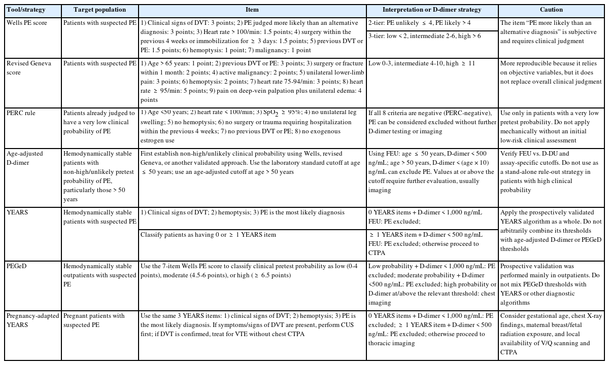
Clinical prediction tools and validated D-dimer strategies for suspected PE and DVT

폐색전증: 혈역학적 안정을 기준으로 한 진단 로드맵

폐색전증이 의심되면 첫 번째 분기점은 혈역학적 안정성이다(Fig. 1의 A). 심정지, 폐쇄성 쇼크, 지속 저혈압 또는 혈압 유지를 위한 승압제 필요가 있으면 고위험 폐색전증을 우선 고려한다[2,4]. 혈역학적으로 불안정한 환자에서는 초기 침상 심초음파가 D-dimer 검사보다 즉각적인 치료 결정에 더 유용하다. 심초음파는 폐색전증을 직접 확진하는 검사는 아니지만 우심실 확장 및 기능 저하, McConnell 징후, 우심방 또는 폐동맥 혈전과 같은 우심실 부담을 확인하고 심낭압전, 급성 좌심실부전, 대동맥질환 등 다른 쇼크 원인을 신속히 감별할 수 있다[2,4]. 환자 이동이 가능하면 폐동맥 컴퓨터단층혈관조영술을 지체 없이 시행하고, 이동이 불가능하면서 임상 가능성이 매우 높고 명확한 우심실 압력 과부하가 확인되면 다른 치명적 원인을 배제하면서 응급 재관류 전략을 판단한다[2,4].
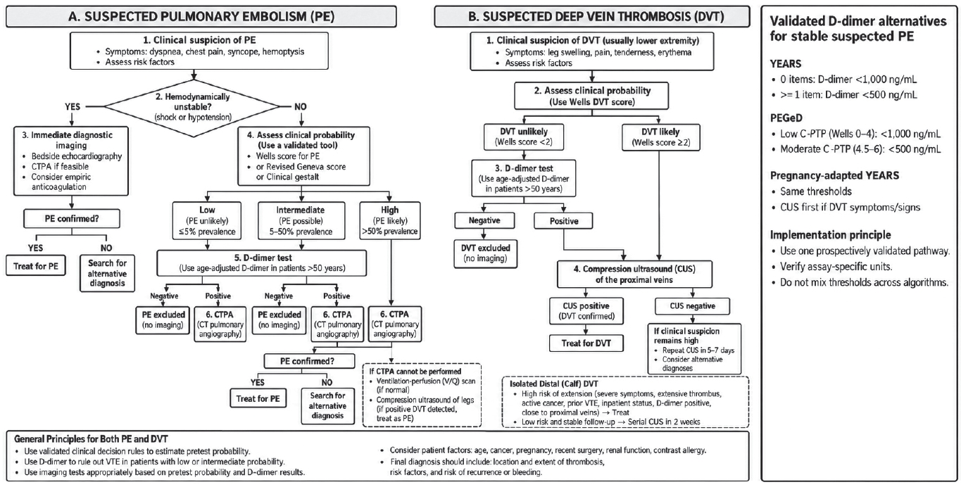
Figure 1.

Diagnostic algorithm for suspected venous thromboembolism. PE, pulmonary embolism; CTPA, computed tomography pulmonary angiography; DVT, deep vein thrombosis; CUS, compression ultrasonography; PEGeD, Pulmonary Embolism Graduated D-dimer; C-PTP, clinical pretest probability; VTE, venous thromboembolism.

혈역학적으로 불안정한 환자에서 D-dimer를 우선하지 않는 주된 이유는 대량 혈전에서 항상 음성이 될 수 있기 때문이 아니라 이미 검사 전 임상 가능성이 높고 즉각적인 해부학적 확인과 위험도 평가가 필요하며 쇼크, 감염, 암, 수술 후 상태 등에서는 D-dimer의 특이도가 낮아 결과가 치료 경로를 바꾸기 어렵기 때문이다[2,4,19]. 또한 임상 가능성이 높은 환자에서 음성 D-dimer만으로 폐색전증을 배제하는 전략은 현재 근거의 정밀도가 충분하지 않으므로 확진 영상 검사를 우선하는 것이 타당하다[2,4,19]. 대부분의 D-dimer 진단 관리 연구가 치료 목적의 항응고를 시작하기 전 환자를 대상으로 했다는 점도 해석에 고려해야 한다[4].

혈역학적으로 안정한 폐색전증 의심 환자에서는 검사 전 임상 가능성에 따라 D-dimer와 영상 검사를 단계적으로 적용한다[2,4]. 낮은 또는 중간 임상 가능성에서 고감도 D-dimer 가 음성이면 추가 영상 없이 폐색전증을 배제할 수 있고, 임상 가능성이 높거나 D-dimer가 양성이면 폐동맥 컴퓨터단층혈관조영술을 시행한다[2,4]. 특히 50세를 초과한 환자에서는 연령 보정 절단값을 사용하면 고정 500 ng/mL 기준보다 불필요한 영상 검사를 안전하게 줄일 수 있다[4,6]. 피브리노겐 등가 단위(fibrinogen-equivalent units, FEU)를 사용하는 경우 일반적으로 연령 × 10 ng/mL를 적용하지만 검사실마다 FEU 와 D-dimer 단위(D-dimer units, D-DU) 등 보고 단위 및 검사법이 다르므로 기관별 기준을 확인해야 한다[4,6]. YEARS 알고리즘은 심부정맥혈전증의 임상 징후, 객혈, 폐색전증이 가장 가능성 높은 진단의 세 항목을 평가하여 항목이 0개이면 D-dimer < 1,000 ng/mL, 1개 이상이면 < 500 ng/mL에서 폐색전증을 배제한다[4,9]. 전향적 YEARS 연구에서는 폐색전증이 배제되어 항응고를 받지 않은 환자의 3개월 진단 실패율이 낮았고 폐동맥 컴퓨터단층혈관조영술 사용을 감소시켰다[9]. 임상 가능성 보정 D-dimer 전략(PEGeD)은 Wells 점수에 따라 낮은 가능성(0-4점)에서 < 1,000 ng/mL, 중간 가능성(4.5-6점)에서 < 500 ng/mL를 사용하여 영상 검사를 줄였다[11]. 2026 미국심장협회/미국심장학회 지침도 임상 가능성이 높지 않은 환자에서 연령 보정 D-dimer 또는 YEARS 기반 전략을 활용하도록 권고한다[4]. 각 알고리즘의 점수 체계와 절단값을 임의로 혼합해서는 안 되며, 대부분의 D-dimer 진단 관리 연구가 치료 목적의 항응고를 시작하기 전 환자를 대상으로 수행되었다는 점도 고려해야 한다[4].

임신 중에는 생리적 변화로 D-dimer가 상승하므로 산모와 태아의 방사선 노출을 함께 고려해야 한다[4,10,20]. 임신 보정 YEARS 알고리즘은 동일한 3가지 YEARS 항목과 D-dimer 절단값을 적용하되 하지 심부정맥혈전증의 증상이나 징후가 있으면 압박초음파를 우선 시행하도록 구성되어 있다[4,10]. 대규모 전향적 연구에서 3개월 진단 실패율은 매우 낮았고 전체 임산부의 약 39%에서 흉부 영상 검사를 피할 수 있었다[10]. 2026 미국심장협회/미국심장학회 지침에서도 임산부에서 임신 보정 YEARS 알고리즘을 이용하여 영상 검사가 필요하지 않은 환자를 선별하는 것이 합리적일 수 있다고 제시한다[4]. 영상 검사가 필요한 경우에는 흉부 X선 소견, 기저 폐질환, 환기-관류 스캔과 폐동맥 컴퓨터단층혈관조영술의 가용성 및 방사선 저감 프로토콜을 함께 고려한다[4,20]. 조영제 알레르기나 중증 신기능 저하로 폐동맥 컴퓨터단층혈관조영술이 곤란하면 환기-관류 스캔과 하지 압박초음파를 대체 전략으로 활용할 수 있다[2,4,20]. 폐색전증이 의심되는 상황에서 압박초음파로 근위부 심부정맥혈전증이 객관적으로 확인되면 흉부 영상 검사가 어렵더라도 정맥혈전색전증으로 치료를 시작할 수 있다[2,4]. 확진된 폐색전증에서 동반 심부정맥혈전증의 발견율은 환자군과 체계적 양측 압박초음파 시행 여부에 따라 달라진다. 국내 단일 기관 코호트에서는 동반 심부정맥혈전증이 45.4%, 근위부 심부정맥혈전증이 39.0%에서 확인되었다[21]. 따라서 동반 심부정맥혈전증의 빈도를 하나의 고정된 수치로 단정하기 보다 검사 프로토콜과 환자군에 따른 차이를 고려하는 것이 타당하다[21].

심부정맥혈전증: D-dimer와 압박초음파의 역할

하지 심부정맥혈전증이 의심되면 Wells 심부정맥혈전증 점수와 같은 임상 예측 도구로 검사 전 가능성을 구분한다(Fig. 1의 B) [1,3,17]. 가능성이 낮은 경우(< 2점) 고감도 D-dimer가 음성이면 영상 검사 없이 심부정맥혈전증을 배제할 수 있다. D-dimer가 양성이거나 가능성이 높은 경우(≥ 2점)에는 근위부 압박초음파를 시행한다[1,3,17,22]. 입원 환자, 암 환자 또는 다른 원인으로 D-dimer가 흔히 상승하는 환자에서는 D-dimer의 효율이 낮으므로 임상 가능성이 높다면 압박초음파를 우선하는 것이 합리적이다[1,3,22].
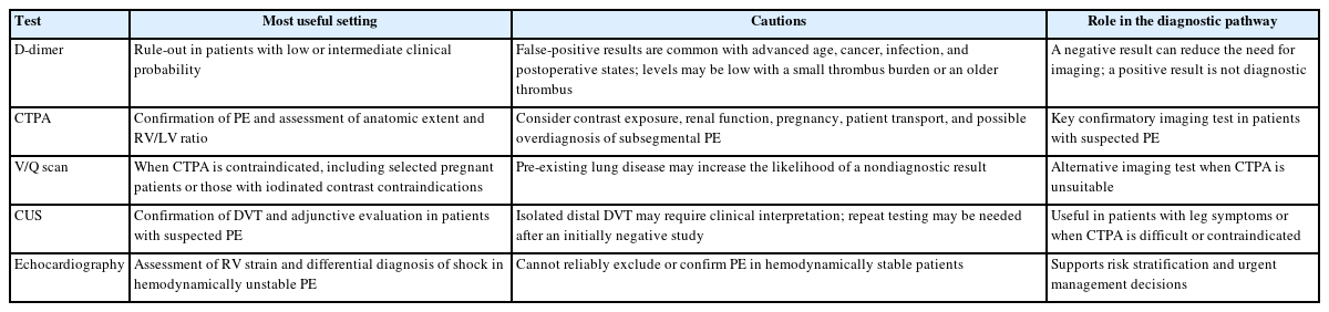
Diagnostic tests and their interpretation in suspected venous thromboembolism

압박초음파에서 정맥이 완전히 압박되지 않는 소견이 심부정맥혈전증의 핵심 진단 기준이다[1,3,5,22]. 근위부 심부정맥혈전증이 확인되면 혈전의 위치와 침범 범위를 명확히 기술하고 즉시 치료로 연결한다. 초기 근위부 압박초음파가 음성이더라도 임상 가능성이 높거나 증상이 지속되면 반복 압박초음파가 필요하며, 임상 프로토콜에 따라 약 1주 이내에 재검한다[1,3,5,22]. 최근 체계적 문헌 고찰에서도 반복 압박초음파는 높은 진단 정확도를 보였다[22]. 전 하지 압박초음파를 한 번에 시행하는 전략도 가능하지만 원위부 혈전의 과잉 진단 가능성을 고려해야 한다[5,22]. 고립성 원위부 심부정맥혈전증에서 증상이 심하거나 활동성 암, 과거 심부정맥혈전증, 광범위 혈전, 근위부 정맥에 가까운 혈전 등 근위부 진행 위험이 높으면 항응고 치료를 우선 고려한다. 반대로 위험이 낮고 추적 관찰이 가능하면 약 1주 간격으로 2주 동안 반복 압박초음파를 시행하는 전략을 사용할 수 있다[5,14]. 최종 보고에는 병변의 측성(좌/우/양측), 침범된 세부 정맥, 근위부/원위부 여부, 혈전의 범위 그리고 급성 또는 만성 소견을 상세히 포함해야 한다.

진단 검사의 해석

D-dimer는 혈전 형성과 섬유소 분해를 반영하는 민감한 검사이지만 특이도가 낮아 낮은 또는 중간 임상 가능성에서 음성일 때 정맥혈전색전증을 배제하는 데 가장 유용하다[1-4]. 반면 고령, 활동성 암, 임신, 감염, 최근 수술이나 외상 및 중증 입원 환자에서는 D-dimer 양성률이 높으므로 양성 결과만으로 정맥혈전색전증을 진단해서는 안 된다[1-4,20]. 특히 임상 가능성이 높은 환자에서는 D-dimer 음성 결과만으로 폐색전증이나 심부정맥혈전증을 배제하는 것이 권장되지 않는다[2,4,19]. 최근 유럽 3개 연구의 사후 분석에서는 높은 임상 가능성을 보인 651명 중 연령 보정 D-dimer가 음성이었던 70명에서 3개월 동안 폐색전증이 관찰되지 않았지만 95% 신뢰구간 상한이 6.5%로 넓어 안전성을 확정하기에는 정밀도가 부족하였다[19]. 따라서 현재 진료 지침에서는 D-dimer 기반 배제 전략을 주로 검사 전 임상 가능성이 높지 않은 환자에게 적용하고, 임상 가능성이 높은 경우에는 폐동맥 컴퓨터단층혈관조영술을 비롯한 확진 영상 검사를 우선하도록 권고한다[2,4,19]. 주요 진단 검사의 역할과 임상적 해석은 표 3에 정리하였다.

폐동맥 컴퓨터단층혈관조영술은 폐색전증의 존재뿐 아니라 주폐동맥, 엽, 분절, 아분절 수준의 해부학적 위치와 우심실 확장, 대체 흉부질환을 평가하는 핵심 검사이다. 다만 조영제 사용, 신기능, 임신, 환자 이동 가능성, 영상의 질 및 아분절폐색전증의 과잉 진단 가능성을 함께 고려해야 한다[2,4,23]. 환기-관류 스캔은 조영제 금기 또는 임신 등에서 대안이 되며[2,4], 압박초음파는 심부정맥혈전증 확진의 핵심 검사이자 폐동맥 컴퓨터단층혈관조영술이 어려운 폐색전증 의심 환자의 보조 진단법이다[1,3-5,22]. 심초음파는 혈역학적으로 안정한 환자에서 폐색전증 자체를 배제하는 검사는 아니지만 불안정한 환자에서 우심실 부담을 신속히 확인하고 확진 후 위험도를 평가하는 데 중요한 정보를 제공한다[2,4].

진단 정보에 기반한 초기 치료 전략

폐색전증이 확진되면 진단 과정에서 확보된 임상 소견과 검사 결과를 바탕으로 초기 치료 장소와 모니터링 강도를 신속히 결정해야 한다[2,4,13,14]. 2019 유럽심장학회 지침에서는 급성 폐색전증의 조기 사망 위험을 저위험, 중등도-저위험, 중등도-고위험 및 고위험의 4단계로 분류한다. 2026 미국심장협회/미국심장학회 지침에서는 급성 폐색전증을 A-E 범주로 새롭게 분류하여 임상적 중증도와 함께 우심실 기능, 심장표지자, 저관류 및 호흡부전의 정도를 보다 세분화하였다[2,4]. 두 분류 체계가 완전히 일대일로 대응하는 것은 아니므로 유럽심장학회 분류를 유지하면서 2026 미국심장협회/미국심장학회 임상 범주를 함께 제시할 수 있다.

혈역학적으로 불안정한 환자는 응급 재관류 치료와 중환자 치료의 필요성을 즉시 평가해야 한다[2,4]. 혈역학적으로 안정하더라도 우심실 기능 저하와 트로포닌 상승이 함께 있는 유럽심장학회 중등도-고위험군 또는 미국심장협회/미국심장학회 C3 범주 환자는 초기 임상 악화 가능성을 고려하여 특히 첫 24-72시간 동안 면밀히 관찰해야 한다[2,4,24]. 반대로 외래 치료 또는 조기 퇴원은 폐색전증 중증도 지수(Pulmonary Embolism Severity Index, PESI), 단순화 폐색전증 중증도 지수(simplified PESI, sPESI), Hestia 기준과 같은 저위험 평가 도구뿐 아니라 산소 요구량, 출혈 위험, 동반 질환 및 사회적 수용 가능성을 함께 고려하여 결정한다[4,7,8,12].

진단 과정에서 확인한 혈역학적 상태, 폐색전증의 위치와 범위, 우심실 기능은 항응고제의 종류나 치료 기간 자체보다는 재관류 치료의 필요성, 초기 치료 장소 및 모니터링 강도를 결정하는 데 더 직접적으로 활용된다[2,4]. 현재 대부분의 진료 지침에서는 혈역학적으로 안정한 심부정맥혈전증과 폐색전증에서 직접 경구항응고제(direct oral anticoagulant, DOAC)를 우선 고려하며[4,13,14], 국내 문헌에서도 새로운 경구항응고제의 실제적 사용과 항응고 치료 기간에 대한 고려사항이 정리된 바 있다[25]. 재관류 치료가 예상되는 고위험 폐색전증에서는 비경구 항응고 전략이 더 적절할 수 있다[2,4]. 영상 정보가 항응고 여부에 직접 영향을 주는 대표적인 예는 고립성 원위부 심부정맥혈전증으로, 근위부 진행 위험이 낮고 신뢰할 수 있는 추적 관찰이 가능하면 즉시 항응고를 시작하는 대신 반복 압박초음파를 선택할 수 있다[5,14]. 반면 항응고제의 구체적인 선택과 치료 기간은 영상에서 보이는 혈전량이나 심초음파의 우심실 기능 저하 자체보다 암, 임신, 삼중 양성 항인지질증후군, 신기능, 출혈 위험, 유발 인자의 지속성 및 재발 위험을 중심으로 개별화해야 한다[3,4,13-15]. 따라서 혈전의 크기나 우심실 기능 저하만을 근거로 항응고 치료 기간을 연장하는 것은 적절하지 않다[4,13,14].

특수 임상 상황에서의 진단적 고려

고령 환자에서는 폐색전증이 전형적인 흉통이나 호흡 곤란보다 실신, 쇠약감, 저산소증, 섬망 또는 심부전 악화로 나타날 수 있다. 또한 D-dimer 양성률이 높으므로 단순한 양성 결과보다 임상 가능성과 영상 소견을 함께 해석하는 것이 중요하다[2,4]. 암 환자는 혈전 위험과 출혈 위험이 모두 높으며 우연히 발견된 폐색전증에서도 혈전의 위치, 증상, 동반 심부정맥혈전증, 암의 활동성, 항암 치료 및 출혈 위험을 종합적으로 평가해야 한다[13-15]. 특히 암 연관 정맥혈전색전증의 항응고 전략은 암종, 장기 기능, 병용 약제 및 출혈 위험에 따라 개별화해야 한다[15].

수술 후 환자에서는 통증, 부종, 빈맥, 산소 요구량 증가가 수술 자체 또는 감염으로 설명되기 쉬워 정맥혈전색전증의 진단이 지연될 수 있다. 그러나 수술 후 새로 발생한 하지부종 또는 설명되지 않는 호흡 곤란이 있으면 심부정맥혈전증과 폐색전증을 함께 평가해야 한다[3]. 임신에서는 임신 보정 YEARS 알고리즘과 증상이 있는 하지의 압박초음파를 우선 고려하고 영상 검사가 필요한 경우에는 산모의 유방과 태아에 대한 방사선 노출, 흉부 X선 소견 및 검사 가용성을 함께 고려한다[4,10,20]. 신기능 저하나 조영제 알레르기가 있는 경우에는 폐동맥 컴퓨터단층혈관조영술의 대체 전략을 미리 정해두는 것이 진단 지연을 줄이는 데 도움이 된다[2,4].

아분절 폐색전증과 고립성 원위부 심부정맥혈전증은 모두 과잉 진단과 과소 치료 사이에서 판단이 어려운 영역이지만 평가 요소는 서로 다르다. 아분절 폐색전증에서는 영상의 질과 진단의 확실성, 단일 또는 다발성 여부, 동반 근위부 심부정맥혈전증, 암, 심폐 예비능, 증상 및 출혈 위험을 종합적으로 평가한다[4,14]. 고립성 원위부 심부정맥혈전증에서는 증상의 정도, 근위부 진행 위험 인자 및 반복 압박초음파 시행 가능 여부를 중심으로 항응고 치료와 영상 추적 중 하나를 선택한다[5,14]. 이러한 경계 영역에서는 확실한 진단과 남아 있는 불확실성을 최종 기록에서 구분하여 이후 치료 및 추적 전략과 연결하는 것이 바람직하다[2,4,5].

추적 관찰 중 진단적 평가의 역할

정맥혈전색전증의 진단은 응급실 또는 입원 첫날의 영상 소견으로 완전히 종료되지 않는다. 항응고 치료 후에도 호흡 곤란이나 하지 증상이 지속되는 환자에서는 폐색전증 후 증후군과 만성 혈전색전성 폐질환(chronic thromboembolic pulmonary disease, CTEPD)을 평가해야 하며[4,26], 만성 혈전색전성 폐고혈압(chronic thromboembolic pulmonary hypertension, CTEPH)은 만성 혈전색전성 폐질환 중 폐고혈압이 동반된 표현형이다[4,26]. 심부정맥혈전증 후에는 만성 부종, 통증 및 피부 변화가 지속될 때 혈전후증후군(post-thrombotic syndrome, PTS)을 고려한다[5,13]. 이는 치료 반응을 평가하는 동시에 초기 진단 당시의 혈전 범위와 중증도를 다시 확인하는 과정이기도 하다[4].

2026 미국심장협회/미국심장학회 지침은 급성 폐색전증 후 최소 1년 동안 매 방문 시 폐색전증 관련 증상과 기능 제한을 확인하도록 권고한다[4]. 특히 적절한 항응고 치료를 3개월 이상 시행했음에도 호흡 곤란 또는 기능 저하가 지속되면 만성 혈전색전성 폐질환에 대한 진단적 평가가 필요하다[4,26]. 만성 혈전색전성 폐질환이 의심되는 경우에는 심초음파 단독보다 심초음파와 폐 관류 영상(평면 환기-관류 영상, 환기-관류 단일광자방출컴퓨터단층촬영 또는 SPECT/CT)을 함께 시행하는 것이 합리적이며 필요에 따라 심폐운동 검사와 추가 폐혈관 평가를 단계적으로 시행한다[4]. 반대로 증상이 소실되고 만성 혈전색전성 폐질환의 의심이 낮은 환자에서는 혈전의 영상학적 소실 여부만 확인하기 위한 정기적 추적 폐동맥 컴퓨터단층혈관조영술 또는 폐 관류 영상 검사는 권장되지 않는다[4].

심부정맥혈전증 후 만성 하지부종, 통증, 피부 변화 또는 정맥성 궤양이 지속되면 혈전후증후군을 평가한다. 잔여 혈전이 관찰된다는 이유만으로 항응고 치료 기간을 자동으로 연장해서는 안 되며, 미국혈액학회 지침에서도 항응고 치료 기간을 결정하기 위해 잔여 정맥 혈전을 확인하는 정기적 초음파 검사를 일률적으로 시행하는 것은 권장하지 않는다[13]. 새로운 하지 증상이 발생하면 재발성 심부정맥혈전증과 만성 변화를 감별하기 위해 초기 또는 이전 영상과 비교하여 선택적으로 압박초음파를 시행한다[5,13]. 따라서 무증상 환자에서 폐동맥 컴퓨터단층혈관조영술 또는 압박초음파를 일률적으로 반복하기보다 증상, 재발 위험 및 치료 결정에 영향을 주는 경우에 선택적으로 영상 검사를 시행하는 것이 타당하다[4,5,13].

증례에 진단 로드맵의 적용

본 증례는 실신과 심한 저산소증을 보인 79세 남자로 검사 전 임상 가능성을 평가한 뒤 폐동맥 컴퓨터단층혈관조영술에서 양측 주폐동맥과 다발성 엽, 분절 폐색전증을 확인하였고 하지 압박초음파에서 양측 슬와정맥의 심부정맥혈전증을 확인하였다. 확진 후 PESI는 109점(IV군), sPESI는 1점이었으며 우심실 수축기능 저하와 고감도 트로포닌 I 상승이 동반되어 2019 유럽심장학회 기준으로 중등도-고위험군에 해당하였다[2]. 또한 혈압은 유지되었지만 젖산이 2.15 mmol/L로 상승해 저관류 소견이 동반되었고, 실내 공기 산소포화도 80%로 호흡 부전이 확인되어 2026 미국심장협회/미국심장학회 임상 범주에서는 D2R 등급으로 분류할 수 있다[4]. 따라서 본 환자는 외래 치료보다 초기 입원과 면밀한 모니터링이 필요한 환자로 판단된다[2,4]. Table 4는 이러한 임상 정보와 위험도 판단을 단계별로 연결하여 진단 로드맵이 실제 임상 의사결정에 어떻게 적용되는지를 보여준다.
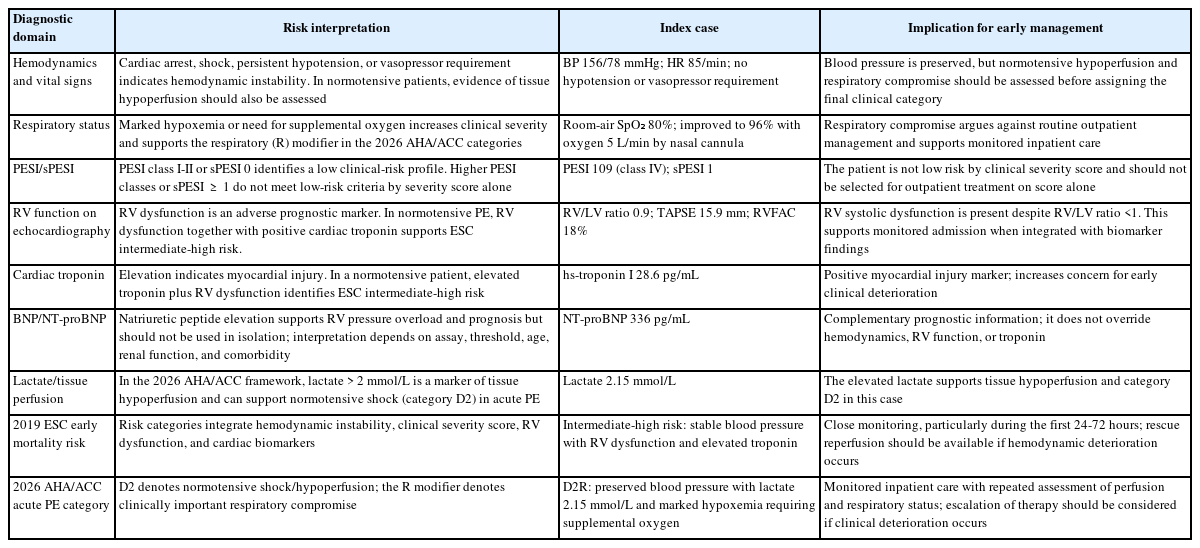
Integration of diagnostic findings with risk stratification and early management in acute pulmonary embolism

결 론

정맥혈전색전증의 진단 로드맵은 임상적 의심과 검사 전 가능성 평가, 폐색전증에서는 혈역학적 안정성, 심부정맥혈전증에서는 하지 임상 가능성에 따른 분기, 검증된 D-dimer 전략과 적절한 영상 검사, 해부학적 확진과 폐색전증의 조기 위험도 분류로 요약할 수 있다. 폐색전증 확진 후에는 기존 유럽심장학회 조기 사망 위험도 분류와 2026 미국심장협회/미국심장학회 급성 폐색전증 임상 범주를 함께 활용하여 우심실 기능 저하, 심장표지자 이상, 저관류 및 호흡부전을 구조적으로 통합해 평가할 수 있다[2,4]. 최종 진단에는 혈전의 위치와 범위, 혈역학적 상태와 우심실 부담, 유발 인자, 재발 위험 및 남아 있는 불확실성을 함께 기록해야 한다. 이후에는 최소 1년 동안 증상과 기능 상태를 확인하고 3개월 이상 증상이 지속되는 경우 만성 혈전색전성 폐질환을 체계적으로 평가하는 추적 진단 경로로 연결하는 것이 중요하다[4].

이와 같은 구조화된 진단 로드맵을 진료 프로토콜과 전자의무기록 의사결정지원에 적용하면 치명적인 정맥혈전색전증의 진단 지연을 줄이는 동시에 임상 가능성이 낮은 환자의 불필요한 영상 검사와 항응고 치료를 줄이고 응급실-입원-외래 추적 사이의 진료 일관성을 높일 수 있다. 향후에는 기관별 진단 실패율, 폐동맥 컴퓨터단층혈관조영술 사용률, 외래 치료 적합성 및 추적 누락을 감사 지표로 연결하여 구조화된 진단 로드맵의 실제 임상 효과를 평가할 필요가 있다.

Notes

CONFLICTS OF INTEREST

No potential conflict of interest relevant to this article was reported.

FUNDING

None.

AUTHOR CONTRIBUTIONS

JS Woo drafted the manuscript, reviewed and revised the manuscript.

ACKNOWLEDGEMENTS

None.

References

1. Lim W, Le Gal G, Bates SM, et al. American Society of Hematology 2018 guidelines for management of venous thromboembolism: diagnosis of venous thromboembolism. Blood Adv 2018;2:3226–3256.
2. Konstantinides SV, Meyer G, Becattini C, et al. 2019 ESC Guidelines for the diagnosis and management of acute pulmonary embolism developed in collaboration with the European Respiratory Society (ERS). Eur Heart J 2020;41:543–603.
3. National Institute for Health and Care Excellence. Venous thromboembolic diseases: diagnosis, management and thrombophilia testing. NICE guideline NG158. London: National Institute for Health and Care Excellence (NICE), 2023.
4. Writing Committee Members, Creager MA, Barnes GD, et al. 2026 AHA/ACC/ACCP/ACEP/CHEST/SCAI/SHM/SIR/SVM/SVN guideline for the evaluation and management of acute pulmonary embolism in adults: a report of the American College of Cardiology/American Heart Association Joint Committee on Clinical Practice Guidelines. Circulation 2026;153:e977–e1051.
5. Mazzolai L, Ageno W, Alatri A, et al. Second consensus document on diagnosis and management of acute deep vein thrombosis: updated document elaborated by the ESC Working Group on aorta and peripheral vascular diseases and the ESC Working Group on pulmonary circulation and right ventricular function. Eur J Prev Cardiol 2022;29:1248–1263.
6. Righini M, Van Es J, Den Exter PL, et al. Age-adjusted D-dimer cutoff levels to rule out pulmonary embolism: the ADJUST-PE study. JAMA 2014;311:1117–1124.
7. Elias A, Mallett S, Daoud-Elias M, Poggi JN, Clarke M. Prognostic models in acute pulmonary embolism: a systematic review and meta-analysis. BMJ Open 2016;6e010324.
8. den Exter PL, Zondag W, Klok FA, et al. Efficacy and safety of outpatient treatment based on the hestia clinical decision rule with or without N-terminal pro-brain natriuretic peptide testing in patients with acute pulmonary embolism. A randomized clinical trial. Am J Respir Crit Care Med 2016;194:998–1006.
9. van der Hulle T, Cheung WY, Kooij S, et al. Simplified diagnostic management of suspected pulmonary embolism (the YEARS study): a prospective, multicentre, cohort study. Lancet 2017;390:289–297.
10. van der Pol LM, Tromeur C, Bistervels IM, et al. Pregnancy-adapted YEARS algorithm for diagnosis of suspected pulmonary embolism. N Engl J Med 2019;380:1139–1149.
11. Kearon C, de Wit K, Parpia S, et al. Diagnosis of pulmonary embolism with D-dimer adjusted to clinical probability. N Engl J Med 2019;381:2125–2134.
12. Roy PM, Penaloza A, Hugli O, et al. Triaging acute pulmonary embolism for home treatment by Hestia or simplified PESI criteria: the HOME-PE randomized trial. Eur Heart J 2021;42:3146–3157.
13. Ortel TL, Neumann I, Ageno W, et al. American Society of Hematology 2020 guidelines for management of venous thromboembolism: treatment of deep vein thrombosis and pulmonary embolism. Blood Adv 2020;4:4693–4738.
14. Stevens SM, Woller SC, Kreuziger LB, et al. Antithrombotic therapy for VTE disease: second update of the CHEST guideline and expert panel report. Chest 2021;160:e545–e608.
15. Kim SA, Yhim HY, Bang SM. Current management of cancer-associated venous thromboembolism: focus on direct oral anticoagulants. J Korean Med Sci 2019;34e52.
16. van Es N, van der Hulle T, van Es J, et al. Wells rule and D-dimer testing to rule out pulmonary embolism: a systematic review and individual-patient data meta-analysis. Ann Intern Med 2016;165:253–261.
17. Kafeza M, Shalhoub J, Salooja N, Bingham L, Spagou K, Davies AH. A systematic review of clinical prediction scores for deep vein thrombosis. Phlebology 2017;32:516–531.
18. Freund Y, Cachanado M, Aubry A, et al. Effect of the pulmonary embolism rule-out criteria on subsequent thromboembolic events among low-risk emergency department patients: the PROPER randomized clinical trial. JAMA 2018;319:559–566.
19. Bannelier H, Kapfer T, Roussel M, et al. Failure rate of D-dimer testing in patients with high clinical probability of pulmonary embolism: ancillary analysis of three European studies. Acad Emerg Med 2025;32:116–122.
20. De Backer J, Haugaa KH, Hasselberg NE, et al. 2025 ESC Guidelines for the management of cardiovascular disease and pregnancy. Eur Heart J 2025;46:4462–4568.
21. Lee JS, Moon T, Kim TH, et al. Deep vein thrombosis in patients with pulmonary embolism: prevalance, clinical significance and outcome. Vasc Specialist Int 2016;32:166–174.
22. Bhatt M, Braun C, Patel P, et al. Diagnosis of deep vein thrombosis of the lower extremity: a systematic review and meta-analysis of test accuracy. Blood Adv 2020;4:1250–1264.
23. Klok FA, Barco S, Bertoletti L, et al. Optimal approach to performing and reporting computed tomography angiography for suspected acute pulmonary embolism: a clinical consensus statement of the ESC working group on Pulmonary Circulation & Right Ventricular Function, the Fleischner Society, the Association for Acute Cardiovascular Care (ACVC) and the European Association of Cardiovascular Imaging (EACVI) of the ESC, Endorsed by European Respiratory Society (ERS), Asian Society of Thoracic Radiology (ASTR), European Society of Thoracic Imaging (ESTI), and Society of Thoracic Radiology (STR). Radiology 2025;315e243833.
24. Meyer G, Vicaut E, Danays T, et al. Fibrinolysis for patients with intermediate-risk pulmonary embolism. N Engl J Med 2014;370:1402–1411.
25. Kim YK. Current management of pulmonary thromboembolism. Korean J Med 2013;84:659–669.
26. Klok FA, Ageno W, Ay C, et al. Optimal follow-up after acute pulmonary embolism: a position paper of the European Society of Cardiology Working Group on Pulmonary Circulation and Right Ventricular Function, in collaboration with the European Society of Cardiology Working Group on Atherosclerosis and Vascular Biology, endorsed by the European Respiratory Society. Eur Heart J 2022;43:183–189.

Article information Continued

Figure 1.

Diagnostic algorithm for suspected venous thromboembolism. PE, pulmonary embolism; CTPA, computed tomography pulmonary angiography; DVT, deep vein thrombosis; CUS, compression ultrasonography; PEGeD, Pulmonary Embolism Graduated D-dimer; C-PTP, clinical pretest probability; VTE, venous thromboembolism.

Table 1.

Clinical clues prompting suspicion of venous thromboembolism

Clinical clue Diagnostic consideration Differential diagnosis Recommended first step
Unilateral leg swelling or pain Consider proximal or distal DVT Cellulitis, lymphedema, chronic venous insufficiency, ruptured Baker cyst Wells DVT score; D-dimer and/or CUS
Bilateral leg swelling Systemic causes are more common; however, bilateral DVT should be considered in patients with cancer, iliac or inferior vena cava thrombosis, or severe immobility Heart failure, renal failure, liver disease, medication-related edema, chronic venous insufficiency Evaluate systemic causes; if VTE risk is high, perform bilateral CUS and consider iliac/inferior vena cava imaging
Dyspnea or chest pain Consider PE and differentiate it from other cardiopulmonary disorders ACS, pneumonia, pneumothorax, aortic disease Assess hemodynamic status; apply Wells PE/revised Geneva assessment; use D-dimer and CTPA as indicated
Syncope or hypoxemia Consider central PE or significant right ventricular strain Arrhythmia, ACS, aortic disease, neurologic causes First assess for high-risk PE and right ventricular dysfunction
Symptoms after surgery, cancer, or prolonged immobility Increased likelihood of provoked VTE Postoperative hematoma, infection, cardiopulmonary complications Assess clinical probability and consider early imaging when indicated

DVT, deep vein thrombosis; CUS, compression ultrasonography; VTE, venous thromboembolism; PE, pulmonary embolism; ACS, acute coronary syndrome; CTPA, computed tomography pulmonary angiography.

Table 2.

Clinical prediction tools and validated D-dimer strategies for suspected PE and DVT

Tool/strategy Target population Item Interpretation or D-dimer strategy Caution
Wells PE score Patients with suspected PE 1) Clinical signs of DVT: 3 points; 2) PE judged more likely than an alternative diagnosis: 3 points; 3) Heart rate > 100/min: 1.5 points; 4) surgery within the previous 4 weeks or immobilization for ≥ 3 days: 1.5 points; 5) previous DVT or PE: 1.5 points; 6) hemoptysis: 1 point; 7) malignancy: 1 point 2-tier: PE unlikely ≤ 4, PE likely > 4 The item “PE more likely than an alternative diagnosis” is subjective and requires clinical judgment
3-tier: low < 2, intermediate 2-6, high > 6
Revised Geneva score Patients with suspected PE 1) Age > 65 years: 1 point; 2) previous DVT or PE: 3 points; 3) surgery or fracture within 1 month: 2 points; 4) active malignancy: 2 points; 5) unilateral lower-limb pain: 3 points; 6) hemoptysis: 2 points; 7) heart rate 75-94/min: 3 points; 8) heart rate ≥ 95/min: 5 points; 9) pain on deep-vein palpation plus unilateral edema: 4 points Low 0-3, intermediate 4-10, high ≥ 11 More reproducible because it relies on objective variables, but it does not replace overall clinical judgment
PERC rule Patients already judged to have a very low clinical probability of PE 1) Age <50 years; 2) heart rate < 100/min; 3) SpO2 ≥ 95%; 4) no unilateral leg swelling; 5) no hemoptysis; 6) no surgery or trauma requiring hospitalization within the previous 4 weeks; 7) no previous DVT or PE; 8) no exogenous estrogen use If all 8 criteria are negative (PERC-negative), PE can be considered excluded without further D-dimer testing or imaging Use only in patients with a very low pretest probability. Do not apply mechanically without an initial low-risk clinical assessment
Age-adjusted D-dimer Hemodynamically stable patients with non-high/unlikely pretest probability of PE, particularly those > 50 years First establish non-high/unlikely clinical probability using Wells, revised Geneva, or another validated approach. Use the laboratory standard cutoff at age ≤ 50 years; use an age-adjusted cutoff at age > 50 years Using FEU: age ≤ 50 years, D-dimer < 500 ng/mL; age > 50 years, D-dimer < (age × 10) ng/mL can exclude PE. Values at or above the cutoff require further evaluation, usually imaging Verify FEU vs. D-DU and assay-specific cutoffs. Do not use as a stand-alone rule-out strategy in patients with high clinical probability
YEARS Hemodynamically stable patients with suspected PE 1) Clinical signs of DVT; 2) hemoptysis; 3) PE is the most likely diagnosis 0 YEARS items + D-dimer < 1,000 ng/mL FEU: PE excluded; Apply the prospectively validated YEARS algorithm as a whole. Do not arbitrarily combine its thresholds with age-adjusted D-dimer or PEGeD thresholds
Classify patients as having 0 or ≥ 1 YEARS item ≥ 1 YEARS item + D-dimer < 500 ng/mL FEU: PE excluded; otherwise proceed to CTPA
PEGeD Hemodynamically stable outpatients with suspected PE Use the 7-item Wells PE score to classify clinical pretest probability as low (0-4 points), moderate (4.5-6 points), or high (≥ 6.5 points) Low probability + D-dimer < 1,000 ng/mL: PE excluded; moderate probability + D-dimer <500 ng/mL: PE excluded; high probability or D-dimer at/above the relevant threshold: chest imaging Prospective validation was performed mainly in outpatients. Do not mix PEGeD thresholds with YEARS or other diagnostic algorithms
Pregnancy-adapted YEARS Pregnant patients with suspected PE Use the same 3 YEARS items: 1) clinical signs of DVT; 2) hemoptysis; 3) PE is the most likely diagnosis. If symptoms/signs of DVT are present, perform CUS first; if DVT is confirmed, treat for VTE without chest CTPA 0 YEARS items + D-dimer < 1,000 ng/mL: PE excluded; ≥ 1 YEARS item + D-dimer < 500 ng/mL: PE excluded; otherwise proceed to thoracic imaging Consider gestational age, chest X-ray findings, maternal breast/fetal radiation exposure, and local availability of V/Q scanning and CTPA

PE, pulmonary embolism; DVT, deep vein thrombosis; PERC, pulmonary embolism rule-out criteria; FEU, fibrinogen-equivalent units; D-DU, D-dimer units; VTE, venous thromboembolism; CTPA, computed tomography pulmonary angiography; V/Q, ventilation-perfusion.

Table 3.

Diagnostic tests and their interpretation in suspected venous thromboembolism

Test Most useful setting Cautions Role in the diagnostic pathway
D-dimer Rule-out in patients with low or intermediate clinical probability False-positive results are common with advanced age, cancer, infection, and postoperative states; levels may be low with a small thrombus burden or an older thrombus A negative result can reduce the need for imaging; a positive result is not diagnostic
CTPA Confirmation of PE and assessment of anatomic extent and RV/LV ratio Consider contrast exposure, renal function, pregnancy, patient transport, and possible overdiagnosis of subsegmental PE Key confirmatory imaging test in patients with suspected PE
V/Q scan When CTPA is contraindicated, including selected pregnant patients or those with iodinated contrast contraindications Pre-existing lung disease may increase the likelihood of a nondiagnostic result Alternative imaging test when CTPA is unsuitable
CUS Confirmation of DVT and adjunctive evaluation in patients with suspected PE Isolated distal DVT may require clinical interpretation; repeat testing may be needed after an initially negative study Useful in patients with leg symptoms or when CTPA is difficult or contraindicated
Echocardiography Assessment of RV strain and differential diagnosis of shock in hemodynamically unstable PE Cannot reliably exclude or confirm PE in hemodynamically stable patients Supports risk stratification and urgent management decisions

CTPA, computed tomography pulmonary angiography; PE, pulmonary embolism; RV/LV, right ventricle/left right ventricle; V/Q, ventilation-perfusion; CUS, compression ultrasonography; DVT, deep vein thrombosis.

Table 4.

Integration of diagnostic findings with risk stratification and early management in acute pulmonary embolism

Diagnostic domain Risk interpretation Index case Implication for early management
Hemodynamics and vital signs Cardiac arrest, shock, persistent hypotension, or vasopressor requirement indicates hemodynamic instability. In normotensive patients, evidence of tissue hypoperfusion should also be assessed BP 156/78 mmHg; HR 85/min; no hypotension or vasopressor requirement Blood pressure is preserved, but normotensive hypoperfusion and respiratory compromise should be assessed before assigning the final clinical category
Respiratory status Marked hypoxemia or need for supplemental oxygen increases clinical severity and supports the respiratory (R) modifier in the 2026 AHA/ACC categories Room-air SpO₂ 80%; improved to 96% with oxygen 5 L/min by nasal cannula Respiratory compromise argues against routine outpatient management and supports monitored inpatient care
PESI/sPESI PESI class I-II or sPESI 0 identifies a low clinical-risk profile. Higher PESI classes or sPESI ≥ 1 do not meet low-risk criteria by severity score alone PESI 109 (class IV); sPESI 1 The patient is not low risk by clinical severity score and should not be selected for outpatient treatment on score alone
RV function on echocardiography RV dysfunction is an adverse prognostic marker. In normotensive PE, RV dysfunction together with positive cardiac troponin supports ESC intermediate-high risk. RV/LV ratio 0.9; TAPSE 15.9 mm; RVFAC 18% RV systolic dysfunction is present despite RV/LV ratio <1. This supports monitored admission when integrated with biomarker findings
Cardiac troponin Elevation indicates myocardial injury. In a normotensive patient, elevated troponin plus RV dysfunction identifies ESC intermediate-high risk hs-troponin I 28.6 pg/mL Positive myocardial injury marker; increases concern for early clinical deterioration
BNP/NT-proBNP Natriuretic peptide elevation supports RV pressure overload and prognosis but should not be used in isolation; interpretation depends on assay, threshold, age, renal function, and comorbidity NT-proBNP 336 pg/mL Complementary prognostic information; it does not override hemodynamics, RV function, or troponin
Lactate/tissue perfusion In the 2026 AHA/ACC framework, lactate > 2 mmol/L is a marker of tissue hypoperfusion and can support normotensive shock (category D2) in acute PE Lactate 2.15 mmol/L The elevated lactate supports tissue hypoperfusion and category D2 in this case
2019 ESC early mortality risk Risk categories integrate hemodynamic instability, clinical severity score, RV dysfunction, and cardiac biomarkers Intermediate-high risk: stable blood pressure with RV dysfunction and elevated troponin Close monitoring, particularly during the first 24-72 hours; rescue reperfusion should be available if hemodynamic deterioration occurs
2026 AHA/ACC acute PE category D2 denotes normotensive shock/hypoperfusion; the R modifier denotes clinically important respiratory compromise D2R: preserved blood pressure with lactate 2.15 mmol/L and marked hypoxemia requiring supplemental oxygen Monitored inpatient care with repeated assessment of perfusion and respiratory status; escalation of therapy should be considered if clinical deterioration occurs

BP, blood pressure; HR, heart rate; AHA/ACC, American Heart Association/American College of Cardiology; PESI, Pulmonary Embolism Severity Index; sPESI, simplified PESI; RV, right ventricle; PE, pulmonary embolism; ESC, European Society of Cardiology; LV, left ventricle; TAPSE, tricuspid annular plane systolic excursion; RVFAC, right ventricular fractional area change; hs-troponin I, high-sensitivity troponin I; NT-proBNP, N-terminal pro-B-type natriuretic peptide; BNP, B-type natriuretic peptide.