피네레논 초기 투여 후 급격한 신기능 악화 증례
Rapid Progression to End-Stage Kidney Disease and Anuria Following Finerenone Initiation in a Patient with Advanced Diabetic Kidney Disease
Article information
Trans Abstract
This case report describes a 70-year-old woman with advanced diabetic kidney disease who developed end-stage kidney disease (ESKD) and complete anuria within seven weeks of initiating finerenone therapy. This case highlights the need for caution when prescribing finerenone to patients with severely impaired renal function, particularly those with minimal residual renal reserve. Clinicians should be vigilant for signs of renal decompensation during the early phases of therapy.
INTRODUCTION
Finerenone, a nonsteroidal mineralocorticoid receptor antagonist (MRA), has demonstrated renal and cardiovascular benefits in patients with type 2 diabetes mellitus (T2DM) and chronic kidney disease (CKD) [1,2]. However, pivotal clinical trials excluded individuals with an estimated glomerular filtration rate (eGFR) of < 25 mL/min/1.73 m2, and the drug’s safety in this population remains uncertain [3].
CASE REPORT
A 70-year-old woman with a 25-year history of poorly controlled T2DM (hemoglobin A1c, 9-11%) and diabetic retinopathy was followed for progressive CKD. Her comorbidities included Parkinson’s disease, which was managed with dopaminergic agents.
Prior to finerenone initiation, laboratory results showed a serum creatinine level of 2.4 mg/dL, an eGFR of 21 mL/min/1.73 m2, a blood urea nitrogen (BUN) of 55.5 mg/dL, and a urine albumin-creatinine ratio (uACR) of 9,344 mg/g creatinine. At the time of finerenone initiation on October 25, 2024, the patient had no fever, infectious symptoms, gastrointestinal illness (vomiting or diarrhea), or documented reduction in oral intake. There was no recent exposure to iodinated contrast agents or nephrotoxic agents (including nonsteroidal anti-inflammatory drugs or herbal medications). The doses of telmisartan and empagliflozin remained unchanged before finerenone initiation, and no intercurrent hypotensive episodes were recorded. The patient was not prescribed any diuretics. Moreover, she had no orthostatic symptoms suggestive of volume depletion and had a stable body weight.
Owing to persistent macroalbuminuria, finerenone 10 mg daily was initiated on October 25, 2024, along with calcium polystyrene sulfonate (one packet twice daily). The patient was also taking empagliflozin 10 mg once daily, pitavastatin 2 mg once daily, telmisartan 40 mg once daily, and premixed insulin (humalog mix 25/75, 16 units twice daily).
Seven weeks later, her renal function rapidly deteriorated. Elevated serum creatinine (7.52 mg/dL), BUN (81.6 mg/dL), and uACR (10,579 mg/g) levels were detected, along with a decrease in eGFR (< 8 mL/min/1.73 m2) and hemoglobin level (8.9 g/dL) (Table 1). Evaluation for other causes of acute kidney injury (AKI) revealed no specific etiology. Viral serologies were also unremarkable (HIV-Ag/Ab-negative, hepatitis B surface antigen [HBsAg]-negative, HCV-Ab-negative, and HBsAb-positive). Autoimmune markers, including ANA, ANCA, anti-glomerular basement membrane antibody, and anti-PLA2R immunoglobulin G (IgG), were negative. Complement levels showed a slightly decreased C3 level (88.8 mg/dL) and an elevated C4 level (49.12 mg/dL). Serum IgG was mildly decreased (376.65 mg/dL), while IgA was within normal limits (231.83 mg/dL). Furthermore, rheumatoid factor levels were low (5.9 IU/mL). Doppler renal ultrasonography revealed diffusely increased renal parenchymal echogenicity in both kidneys, consistent with renal disease. The right kidney measured 8.68 cm and the left kidney 9.18 cm in length, with cortical thicknesses of 0.91 cm (right) and 1.05 cm (left), respectively. Evaluation of the renal arteries was incomplete because of obesity. Therefore, non-contrast abdominopelvic computed tomography was performed and revealed bilateral perinephric infiltration/fluid, which was interpreted as being consistent with AKI.
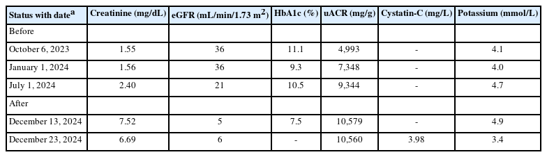
Renal function, glycemic control, and proteinuria before and after finerenone initiation
Baseline transthoracic echocardiography demonstrated left atrial enlargement (left atrium volume index, 38.8 mL/m2) with concentric left ventricular hypertrophy (left ventricle mass index, 123 g/m2; relative wall thickness, 0.44) and papillary muscle hypertrophy with accelerated left ventricle outflow tract flow. Left ventricular systolic function was preserved (ejection fraction, 71%) without regional wall motion abnormalities, but there was grade 2 diastolic dysfunction with impaired relaxation and elevated left atrial pressure (average E/e′, 16.8). Valvular assessment revealed mild degenerative changes (aortic valve calcification at the non-coronary cusp and mild posterior mitral annular calcification) without significant valvular stenosis or regurgitation. Borderline pulmonary hypertension (right ventricular systolic pressure, 38 mmHg) without inferior vena cava plethora or pericardial effusion was observed. During clinical deterioration, the N-terminal pro-B-type natriuretic peptide level was markedly elevated (> 35,000 pg/mL), supporting significant volume overload against a background of diastolic dysfunction and chronically increased filling pressures. These findings indicated that the patient had preserved left ventricular ejection fraction with structural heart disease and elevated cardiac filling pressures, which contributed to the rationale for initiating finerenone despite her advanced diabetic CKD, albuminuria, and high cardiovascular risk.
Blood cultures, both aerobic and anaerobic, showed no growth after 5 days. Serum and urine protein electrophoresis revealed albumin-dominant polyclonal gammopathy, whereas immunofixation electrophoresis showed no evidence of monoclonal gammopathy.
The patient presented with profound clinical deterioration, characterized by complete anuria, dizziness, and severely elevated blood pressure (200/90 mmHg). Arterial blood gas analysis revealed metabolic acidosis (pH, 7.28; PaCO2, 32 mmHg; PaO2, 88 mmHg; and HCO3-, 15.1 mmol/L). Chest radiography revealed signs of volume overload, including mild cardiomegaly, bilateral pleural effusion, and moderate pericardial effusion. Emergency hemodialysis was initiated, and a permanent arteriovenous fistula was created. No reversible causes of AKI such as hypotension, sepsis, or nephrotoxin exposure were identified. The patient remained dialysis-dependent.
DISCUSSION
This case suggests that in patients with an eGFR of < 25 mL/min/1.73 m2 and minimal residual renal reserve, the addition of finerenone to ongoing treatment with renin-angiotensin-aldosterone system (RAAS) inhibition and sodium-glucose cotransporter 2 (SGLT2) blockade may be followed by severe renal decompensation. This likely reflects multifactorial hemodynamic vulnerability rather than finerenone-specific nephrotoxicity. Although a transient decline in eGFR is anticipated with MRA or SGLT2 inhibitor therapy owing to hemodynamic shifts [2,4-6], irreversible renal failure and anuria are rare.
The FIDELIO-DKD trial demonstrated the efficacy of finerenone in reducing the risk of end-stage kidney disease (ESKD), but it notably excluded patients with an eGFR of < 25 mL/min/1.73 m2 [1]. Therefore, the safety of finerenone in patients with advanced kidney dysfunction remains uncertain, and its use in this population should be approached with caution. Reversible AKI shortly after finerenone initiation has been reported in one case report, with improvement after drug discontinuation and supportive care [7].
Our patient was likely vulnerable because of her advanced CKD and severely reduced renal reserve at baseline (eGFR, 21 mL/min/1.73 m2). Although finerenone is known to effectively reduce albuminuria and cardiovascular risk in patients with T2DM and CKD, its administration may pose significant risks, including hyperkalemia, acute renal decompensation, and hypovolemia-related AKI, particularly when used concurrently with SGLT2 inhibitors or in patients with underlying volume depletion [3,8-10].
In our patient, serum potassium levels remained within the normal range throughout the observation period, without any documented episodes of hyperkalemia. The values were as follows: 4.1 mmol/L on October 6, 2023, 4.0 mmol/L on January 1, 2024, and 4.7 mmol/L on July 1, 2024, all before finerenone initiation; and 4.9 mmol/L on December 13, 2024 and 3.4 mmol/L on December 23, 2024, after finerenone initiation (Table 1). Anuria developed between the latter two time points (December 13, 2024 and December 23, 2024). However, the patient’s potassium level was not elevated, either before or during the onset of renal function deterioration. These data indicate that hyperkalemia did not precede or coincide with the development of anuria in our patient. This observation supports our interpretation that rapid kidney decompensation was not mediated by finerenone-induced hyperkalemia, although hyperkalemia remains the most common and clinically relevant adverse event associated with MRAs.
Sarcopenia may lead to the overestimation of kidney function when measuring creatinine-based eGFR in older patients with comorbid conditions such as Parkinson’s disease. According to the KDIGO guidelines, cystatin C or combined creatinine-cystatin C equations are recommended when muscle mass abnormalities may affect eGFR accuracy [11]. However, in the present case, several findings argued against profound creatinine suppression due to severe sarcopenia. Immediately before dialysis initiation, serum creatinine was 7.52 mg/dL, and cystatin C was 3.98 mg/L, yielding a creatinine-to-cystatin C ratio of approximately 1.9 and a cystatin C/creatinine ratio of 0.53. Prior studies have shown that low creatinine-to-cystatin C ratios or high cystatin C/creatinine ratios are associated with sarcopenia and low skeletal muscle mass [12-14], whereas higher creatinine-to-cystatin C ratios generally indicate preserved muscle mass and strength [12,13]. Furthermore, when the CKD-EPI cystatin C equation was applied to the pre-dialysis cystatin C value, the cystatin C-based eGFR fell within the same advanced CKD range as the creatinine-based estimate at that time [11]. Collectively, these biomarker findings suggest that the baseline creatinine-based eGFR of 21 mL/min/1.73 m2 likely reflected advanced but not yet end-stage diabetic kidney disease rather than frank kidney failure at the time of finerenone initiation. Nonetheless, the absence of cystatin C measurements remains an important limitation. Therefore, cystatin C levels should be considered when interpreting the true degree of renal impairment during drug initiation.
Our patient experienced a rapid and irreversible decline in kidney function, culminating in anuric AKI and emergency hemodialysis within 7 weeks of initiating finerenone treatment. Importantly, there were no identifiable reversible causes such as hypotension, nephrotoxins, or infections. From a causal perspective, this event should be interpreted in the context of the patient’s preexisting trajectory of advanced diabetic kidney disease (DKD) and combined hemodynamic stressors. As shown in Table 1, the patient’s serum creatinine increased from 1.55 mg/dL (eGFR, 36 mL/min/1.73 m2) in October 2023 to 2.4 mg/dL (eGFR, 21 mL/min/1.73 m2) in July 2024, indicating progressive stage 4 CKD and markedly reduced renal reserve before finerenone initiation. In addition, the concomitant use of telmisartan, empagliflozin, and finerenone in an older patient with frailty can increase the risk of hemodynamic AKI. Therefore, we do not claim that this case represents finerenone-specific toxic nephropathy. Rather, we consider this episode to be a probable multifactorial hemodynamic collapse occurring in close temporal association with the introduction of finerenone in the setting of aggressive RAAS and SGLT2 inhibition in a patient with minimal residual kidney function. The 7-week interval between finerenone initiation and repeat laboratory testing represents an important limitation and may have delayed the recognition of early renal decline. In similar high-risk patients with an eGFR of < 25 mL/min/1.73 m2, serum creatinine, eGFR, and potassium should be rechecked within 1-2 weeks of starting finerenone and at short intervals thereafter. Although we cannot fully separate drug-related effects from the natural progression of advanced DKD, this case highlights a clinically relevant scenario in which triple blockade in a fragile patient was followed by a rapid, irreversible transition to dialysis-dependent ESKD. Based on this case, cautious use and strict early monitoring of finerenone in this population are essential.
To the best of our knowledge, no previous case has documented such a rapid progression to dialysis-dependent ESKD with anuria in a patient with diabetic nephropathy shortly after initiating finerenone therapy. This case highlights the importance of careful patient selection and monitoring when finerenone is prescribed. In addition, patients with severely reduced kidney function should be carefully monitored if finerenone is prescribed. Guideline-based contraindications should be strictly observed, and the benefits of MRA therapy should be carefully weighed against the potential for acute renal hazards in high-risk populations.
CONCLUSION
Finerenone should be prescribed with extreme caution in patients with severely reduced eGFR, and these patients should be closely monitored during the initial treatment phase to prevent potentially irreversible renal injury.
Notes
CONFLICTS OF INTEREST
The authors declare no conflicts of interest.
FUNDING
None.
AUTHOR CONTRIBUTIONS
Jung Soon Kim: conceptualization, investigation, formal analysis, writing - original draft, writing - review & editing; Hong Il Lim: conceptualization, investigation, formal analysis, writing - original draft, writing - review & editing; So Young Lee: writing - review & editing; Hyo-Jeong Kim: writing - review & editing; Kyong Yeun Jung: writing - review & editing; Jin Taek Kim: conceptualization, investigation, formal analysis, writing - original draft, writing - review & editing.
ACKNOWLEDGEMENTS
We thank Hye Young Han from Eulji University Hospital Library for manuscript’s technical editing.