피마사르탄 재복용 및 간 생검으로 확인된 약인성 간 손상 1예
A Case of Fimasartan-Induced Liver Injury Proven by Rechallenge and Biopsy
Article information
Trans Abstract
Fimasartan is a widely used angiotensin II receptor blocker (ARB) known for its favorable safety profile. However, its hepatotoxic potential remains poorly characterized. We report a case of drug-induced liver injury (DILI) in a 79-year-old woman who developed jaundice and elevated liver enzymes after initiating fimasartan. Liver biopsy revealed severe lobular hepatitis with confluent necrosis. Liver function normalized following drug discontinuation and corticosteroid therapy; however, the injury recurred after accidental reexposure to fimasartan and resolved again upon withdrawal. This temporal association and recurrence upon rechallenge allowed for a definitive diagnosis, with a Roussel Uclaf causality assessment method score of 12 indicating “highly probable” causality. Although ARBassociated DILI is uncommon, this case highlights the need for clinicians to remain vigilant regarding the potential hepatotoxicity of fimasartan. Clinicians should monitor liver function closely during treatment and consider alternative agents if injury occurs.
INTRODUCTION
Fimasartan is an angiotensin II receptor blocker (ARB) that selectively binds to the angiotensin II type 1 receptor, providing a potent antihypertensive effect and a long duration of action. The drug is widely prescribed in Korea and other Asian countries due to its favorable safety profile and low incidence of adverse effects [1]. Liver enzyme elevations associated with fimasartan are rare (less than 2% of patients) and are mostly transient [2]. However, these elevations can occasionally progress to drug-induced liver injury (DILI) characterized by severe liver enzyme elevation. To date, few case reports have demonstrated a clear causal relationship between fimasartan and DILI. Therefore, we aimed to investigate the risk of fimasartan-induced hepatotoxicity through a case in which the causal relationship between fimasartan and DILI has been established.
CASE REPORT
A 79-year-old female patient was referred to our hospital with a 2-week history of nausea and dyspepsia, and a 2-kg weight loss over the preceding month. She had a 20-year history of hypertension and hyperlipidemia, and an 1-year history of prediabetes. Five months earlier, her antihypertensive drug was switched to fimasartan. Her other long-term medications included rosuvastatin (5 mg) and ezetimibe (10 mg), which she had taken for several years. She denied alcohol consumption or the use of herbal medications/supplements. Physical examination revealed generalized jaundice.
Initial serum biochemistry tests showed elevated levels of aspartate aminotransferase (AST; 1,136 IU/L), alanine aminotransferase (ALT; 1,108 IU/L), alkaline phosphatase (ALP; 198 IU/L), gamma-glutamyl transpeptidase (657 IU/L), total bilirubin (3.67 mg/dL), direct bilirubin (1.92 mg/dL), and lactate dehydrogenase (1,137 IU/L). Serological tests for viral hepatitis were all negative for anti-HAV IgM, HBsAg, anti-HBs, anti-HCV, and anti-HIV. Autoimmune antibodies were negative for anti-smooth muscle antibody, anti-liver kidney microsomal type 1 (LKM-1) antibody, and anti-mitochondrial antibody. However, anti-nuclear antibody was positive at a titer of 1:160. Serum immunoglobulin G (IgG) was elevated at 1,785 mg/dL. Chest and contrast-enhanced abdominal computed tomography showed no remarkable findings. Abdominal ultrasound and magnetic resonance cholangiopancreatography revealed no dilation of the main pancreatic duct or bile ducts. A liver biopsy demonstrated severe lobular hepatitis with confluent necrosis, predominantly in the centrilobular area (Fig. 1).
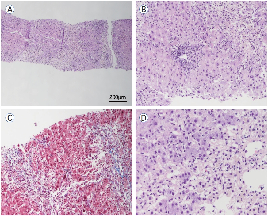
(A) Liver biopsy revealed confluent necrosis of hepatocytes at low power view (HE, × 40). (B, C) Centrilobular necrosis and regenerative hepatocytes in the periportal area were observed on HE and trichrome staining (× 100). (D) Severe hepatocyte apoptosis was identified in zone 3 (HE, × 400). HE, hematoxylin and eosin.
Given the suspicion of DILI or autoimmune hepatitis, all medications were discontinued on hospital day 5, and methylprednisolone 40 mg/day was initiated intravenously. Subsequently, the levels of liver enzymes and bilirubin steadily declined, and the patient was discharged on day 8 in a stable, asymptomatic condition. Three weeks after completing an 1-month gradual steroid taper, follow-up laboratory test con-firmed near-complete resolution: AST 45 IU/L, ALT 11 IU/L, and ALP 99 IU/L.
Two months after discontinuation of steroid therapy, the patient presented to the outpatient clinic with recurrent jaundice. It was subsequently discovered that the patient had accidentally restarted fimasartan 2 weeks prior to the visit. On presentation, repeat serum biochemistry tests showed AST 869 IU/L, ALT 122 IU/L, and ALP 167 IU/L. The patient was immediately readmitted, fimasartan was discontinued, and prednisolone 30 mg/day was administered orally. Subsequently, liver enzyme levels gradually decreased, and she was discharged on day 5 of hospitalization. After discharge, prednisolone was tapered by 5 mg per week. The patient is currently receiving S-amlodipine 5 mg and carvedilol 12.5 mg for hypertension, with no further liver enzyme elevation observed. The changes in the patient’s liver enzyme levels over time are illustrated in Figure 2.
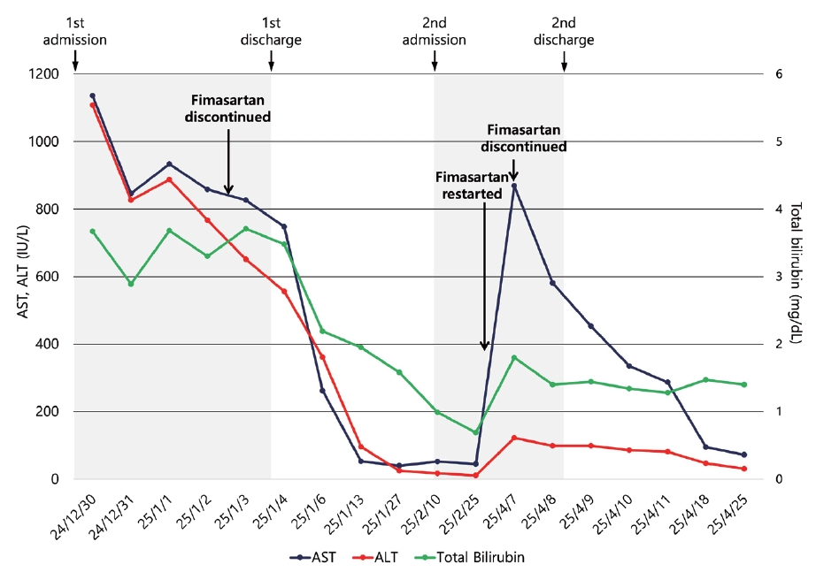
The time-course of liver enzyme levels showed elevation following fimasartan administration, improvement after discontinuation, and re-elevation upon re-exposure. AST, aspartate aminotransferase; ALT, alanine aminotransferase.
Based on the time point after drug re-exposure, the modified Roussel Uclaf causality assessment method (RUCAM) score was 12, indicating a “highly probable” DILI. Key components of the score included: time to onset within 14 days upon re-exposure (+ 2), rapid ALT decline of ≥ 50% within 8 days (+ 3), age ≥ 55 years (+ 1), exclusion of alternative causes (+ 1), hepatotoxicity labeled in the product (+ 2), and ALT doubling upon re-administration (+ 3).
This study was approved by the Institutional Review Board (IRB) of Soonchunhyang University Bucheon Hospital (IRB No. SCHBC 2025-08-004).
DISCUSSION
Fimasartan is an ARB developed in South Korea and approved by the Ministry of Food and Drug Safety in 2010. It was synthesized by replacing the imidazole ring of losartan with a pyrimidin-4(3H)-one ring. Fimasartan is predominantly excreted via the bile, with a lower proportion eliminated in the urine compared to other ARBs [3]. It is currently approved for use mainly in Asian markets such as China, Singapore, and Russia, but is not yet marketed in the United States or European Union.
The understanding of fimasartan-induced DILI remains incomplete. A retrospective cohort study using electronic health records in Korea reported an incidence rate of ARB-induced DILI (defined as ALT ≥ 5× upper limit of normal [ULN] or ALP ≥ 2× ULN) of 48.4 cases per 1,000 person-years, with the majority of cases categorized as mild [4]. This study found no significant differences in DILI risk among the various ARBs evaluated. Conversely, another hospital-based retrospective cohort study demonstrated that fimasartan was associated with a significantly higher frequency of ALT elevations and relatively greater proportion of DILI cases with RUCAM scores ≥ 6, compared with other ARBs [2].
Although numerous case reports have described DILI associated with other ARBs, such as losartan, irbesartan, candesartan, and valsartan, reports specifically implicating fimasartan remain relatively scarce [5,6]. Park et al. [7] reported the first known case of fimasartan-induced DILI, which presented a RUCAM score of 11, indicating “highly probable” causality. Similarly, we previously described a case of fimasartan-associated DILI with a RUCAM score of 6 [8]. Furthermore, the Health Sciences Authority of Singapore reported four cases of fimasartan-related liver injury within a single year [9]. These cases varied in severity; notably, all patients achieved full recovery following drug discontinuation.
Establishing a definitive diagnosis of DILI can be challenging due to the ethical and clinical constraints surrounding intentional drug rechallenge. In the present case, however, the causal relationship between fimasartan and liver injury was supported by several key findings. First, liver enzyme levels increased again following the accidental re-administration of fimasartan, demonstrating a definitive temporal relationship and consistency of association. Second, alternative causes of liver injury were systematically excluded.
Initially, the possibility of autoimmune hepatitis (AIH) could not be ruled out due to elevated antinuclear antibody titers and serum IgG levels. When assessed using the simplified International Autoimmune Hepatitis Group (IAIHG) scoring system, the patient reached a total score of 6 points, categorized as “probable AIH.” However, DILI can frequently manifest with similar immunologic features, thereby mimicking AIH, a condition often referred to as drug-induced autoimmune-like hepatitis. In such cases, the clinical course following corticosteroid withdrawal serves as a primary discriminating factor; while idiopathic AIH typically relapses after treatment cessation, DILI-related injury does not.
In our case, liver enzyme levels remained stable 1 month after the initial drug withdrawal, and no relapse occurred during the 6-month follow-up period following the second episode. Furthermore, the normalization and subsequent stability of serum IgG levels within one month of the second discontinuation strongly support the absence of an underlying chronic autoimmune process. Importantly, the histological evaluation provided pivotal evidence favoring DILI over AIH. The biopsy revealed predominant centrilobular inflammation, whereas hallmark features of AIH such as dense plasma cell infiltration or significant interface hepatitis were notably absent. Taken together, the clinical and histological findings support fimasartan-induced DILI as the primary diagnosis.
Another noteworthy aspect of our case is the latency period. In this instance, DILI developed approximately 5 months after drug initiation. Although this duration is consistent with previously reported fimasartan cases (10 and 7 months, respectively) [7,8], it appears relatively prolonged compared with cases involving other ARBs. Notably, a prior study reported that fimasartan is associated with a higher proportion of patients developing DILI more than 60 days after the initial prescription compared with other drugs in the same class [4]. Given this potential for delayed onset, clinicians must maintain a high index of suspicion for late-developing hepatotoxicity when prescribing fimasartan.
In contrast, the latency period during the second episode was significantly shorter, lasting approximately 2 weeks. This rapid recurrence is highly suggestive of an immune-mediated idiosyncratic reaction, distinguishing it from direct, dose-dependent toxicity. It is highly probable that previously sensitized memory T cells triggered a more vigorous response upon re-exposure to fimasartan, driving rapid clinical progression observed during the second episode. While it is not yet fully established whether DILI results exclusively from immune mechanisms, the appropriateness of corticosteroid therapy also remains controversial. However, there is growing theoretical support for its use; specifically, adaptive immune mechanisms involving CD8+ T cells and mononuclear phagocytes have been linked to DILI pathogenesis [10]. Further prospective studies are warranted to establish clear, evidence-based guidelines for corticosteroid implementation in DILI.
In conclusion, this case provides definitive evidence of fimasartan-induced DILI, confirmed by both positive drug rechallenge and histological findings. Given the expanding clinical use of fimasartan and the limited data on its hepatotoxicity, clinicians should assess baseline liver function prior to treatment initiation and perform routine biochemical monitoring during the initial months of therapy. Further research and accumulation of clinical cases are warranted to better characterize the predisposing risk factors and underlying mechanisms associated with fimasartan-induced hepatotoxicity.
Notes
CONFLICTS OF INTEREST
Authors have no conflict of interests.
FUNDING
The study was supported by Soonchunhyang University Research Fund (2025). The funders had no role in study design, data collection and analysis, decision to publish, or preparation of the manuscript.
AUTHOR CONTRIBUTIONS
Seryeong Kim: data curation, formal analysis, investigation, visualization, writing - original draft, writing - review & editing.
Jisoo Lee: formal analysis, investigation, writing - review & editing.
Susie Chin: resources, writing - review & editing.
Sang Gyune Kim: writing - review & editing, supervision.
Young-Seok Kim: writing - review & editing, supervision.
Jeong-Ju Yoo: conceptualization, funding acquisition, validation, writing - review & editing.
ACKNOWLEDGEMENTS
None.