


종양으로 오인될 수 있는 결절성 근육 유육종증 1예
- 안세한, 김민수, 한민성, 윤정호, 국은희, 전세용, 김철현
A Case of Nodular Muscular Sarcoidosis Mimicking a Tumor
- Se Han Ahn, Min Su Kim, Min Sung Han, Jung Ho Yoon, Eun Hee Kook, Se Yong Jeon, Cheol Hyeon Kim
- Received August 12, 2008; Revised September 8, 2008; Accepted September 29, 2008;
- Abstract
-
The nodular form of muscular sarcoidosis is a rare malady that is often confused with a soft-tissue neoplasm or other lesion. Here, we present a case of nodular muscular sarcoidosis in the arms and legs of a 59-year-old woman. She presented at our hospital with a painless nodule in her left arm. Excision was performed and she was diagnosed with sarcoidosis. One year later, nodular sarcoidosis recurred in her arms and legs. After 2 months of steroid medication, the nodules disappeared. The patient has been followed for 2 years and no evidence of recurrence has been observed. (Korean J Med 2011;80:247-250)
- INTRODUCTION
- INTRODUCTION
Sarcoidosis is a multisystemic granulomatous disease of unknown cause. The lungs, skin, eyes, liver, and lymph nodes are commonly affected, but muscular sarcoidosis is rare. Muscular involvement in sarcoidosis was first reported by Licharew in 1908 [1]. The symptomatic involvement of muscles occurs much less frequently than does asymptomatic involvement [1,2]. Three main clinical types of sarcoidosis have been reported for symptomatic muscular sarcoidosis: nodular, acute myositic, and chronic myopathic. The palpable nodular type reported by Licharew is the least common clinical type [3]. The nodular type can present with a soft-tissue mass that may be confused with a tumor, and magnetic resonance imaging (MRI) and muscle biopsy are useful methods for early diagnosis. The optimal management of the nodular type of muscular sarcoidosis is not well known, but surgical excision or corticosteroid treatment are initial options [4]. We report here the case of a woman with recurrent nodular muscular sarcoidosis who was treated with steroids.
- CASE REPORT
- CASE REPORT
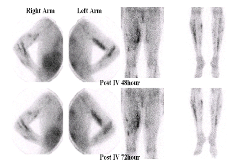
A 58-year-old Korean woman presented with a painless mass in her left arm. A large, firm, fixed mass was palpable in the biceps muscle. Radiographs of the arm revealed a soft-tissue shadow in the biceps. MRI demonstrated that the mass (18×2.3×1.6 cm) had irregular margins (Fig. 1) and enhanced heterogeneously with various signal intensities. The characteristics of this infiltrating mass were most consistent with a soft-tissue sarcoma or fibromatosis. An incision biopsy demonstrated chronic granulomatous inflammation with focal necrosis, suggesting sarcoidosis. Excision was thus performed.One year after excision, the patient returned to our hospital with multiple painless, mass-like lesions in her arms and legs. Ultrasonography demonstrated hypoechoic masses in the right upper arm (11×0.6×1.2 cm), left forearm (8.5×1×1 cm), right lower leg (6.3×1.2×2.4 cm), and right calf (13×1.5×2.6 cm). A gun biopsy was performed on the right leg mass. The biopsy revealed chronic granulomatous inflammation, compatible with sarcoidosis (Fig. 2). Acid-fast staining and Gomori’s methenamine-silver staining showed no microorganism. The chest radiograph was not remarkable, but the chest computed tomography image revealed bilateral hilar lymphadenopathy, consistent with stage I sarcoidosis (Fig. 3). Additional laboratory tests that measured blood cell count, serum angiotensin-converting enzyme level, serum electrolytes, serum calcium level, erythrocyte sedimentation rate, and C-reactive protein level were all within normal limits. The level of creatinine phospho-kinase and the creatine kinase-myocardial band were normal. Gallium uptake was detected in the upper and lower extremities by 67Ga-scintigraphy (Fig. 4). Corticosteroid therapy (prednisolone: 1 mg/kg/day) was initiated and the masses gradually decreased in size during the following week. The prednisolone dose was tapered until treatment was discontinued at 2 months. No sign of recurrence has been detected during 2 years of follow-up.
- DISCUSSION
- DISCUSSION
Asymptomatic muscle involvement occurs in 50~80% of sarcoidosis patients, but symptomatic involvement is rare [1,2]. Three forms of symptomatic muscular sarcoidosis have been described: nodular involvement, acute sarcoid myositis, and chronic sarcoid myopathy [5]. The nodular type is the least common type of symptomatic muscle involvement in sarcoidosis [3]. Although any skeletal muscle may be involved, the lower limbs are most frequently affected. The nodules are painless and are not accompanied by any limitation of movement or muscle weakness. However, contracture or myalgia may occur [5]. Serum creatine kinase levels and electromyographic results are typically normal. The excellent tissue contrast of MRI allows the detection of small nodules, and the MRI findings of the nodular type of sarcoidosis have been reported to be specific [6]. The cytological differential diagnosis of nodular muscular sarcoidosis includes tuberculosis, foreign-body granuloma, fungal or parasitic infection, and autoimmune diseases, such as rheumatoid arthritis [7].Patients with nodular lesions and acute myositis can usually be effectively treated with corticosteroids at an initial dosage of 0.5~1 mg/kg/day [8,9]. Early initiation of treatment appears to increase its effectiveness. Although corticosteroid treatment was found to be rapidly effective for patients with nodular muscular sarcoidosis [9], relapse was observed in all patients several years later. The response to steroids is unpredictable in patients with the chronic myopathic type of muscular sarcoidosis.Patient outcomes may be complicated by corticosteroid-induced myopathy [7]. Chloroquine, azathioprine, and methotrexate should be administered to steroid-dependent patients or those who fail to respond to corticosteroids [3,8,10]. New agents, including thalidomide and infliximab, may also be useful in selected cases [10]. Physicians should consider this unusual presentation of sarcoidosis and its responsiveness to steroid treatment when managing sarcoidosis patients.
- Acknowledgments
- Acknowledgments
We report here the case of a woman with recurrent nodular muscular sarcoidosis who was treated with steroids. Physicians should consider this unusual presentation of sarcoidosis and its responsiveness to steroid treatment when managing sarcoidosis patients.
Figure 1.
Magnetic resonance image showing a heterogeneously enhanced mass (arrow) with various signal intensity components in the anterior arm, involving the biceps muscle. No bony abnormality is visible.

Figure 2.
The biopsy specimen of the soft-tissue mass consisted of confluent nodules of noncaseating granulomatous inflammation (arrow; H&E, ×100).

Figure 3.
Chest computed tomography image showing nonspecific lymphadenopathy in the bilateral hilar areas.

Figure 4.
Radioisotope scanning was performed 48 and 72h after injection of 67Ga. Gallium uptake was detected in all upper and lower extremities.
- References
- References
REFERENCES
1. Silverstein A, Siltzbach LE. Muscle involvement in sarcoidosis: asymptomatic, myositis, and myopathy. Arch Neurol 1969;21:235–241.
[Article] [PubMed]2. Prayson RA. Granulomatous myositis: clinicopathologic study of 12 cases. Am J Clin Pathol 1999;112:63–68.
[PubMed]3. Zisman DA, Biermann JS, Martinez FJ, Devaney KO, Lynch JP 3rd. Sarcoidosis presenting as a tumor like muscular lesion. Case report and review of the literature. Medicine 1999;78:112–122.
[Article] [PubMed]4. Cameron HU. Symmetrical muscle contractures in tumorous sarcoidosis: report of a case. Clin Orthop Relat Res 1981;155:108–110.
[Article] [PubMed]5. Guo M, Lemos L, Baliga M. Nodular sarcoid myositis of skeletal muscle diagnosed by fine needle aspiration biopsy: a case report. Acta cytol 1999;43:1171–1176.
[Article] [PubMed]6. Guis S, Mattéi JP, Lioté F. Drug-induced and toxic myopathies. Best Pract Res Clin Rheumatol 2003;17:877–907.
[Article] [PubMed]7. Tambouret R, Geisinger KR, Powers CN, et al. The clinical application and cost analysis of fine-needle aspiration biopsy in the diagnosis of mass lesions in sarcoidosis. Chest 2000;117:1004–1011.
[Article] [PubMed]8. Sève P, Zénone T, Durieu I, Pillon D, Durand DV. Muscular sarcoidosis: apropos of a case. Rev Med Interne 1997;18:984–988.
[Article] [PubMed]9. Fayad F, Lioté F, Berenbaum F, Orcel P, Bardin T. Muscle involvement in sarcoidosis: a retrospective and followup studies. J Rheumatol 2006;33:98–103.
[PubMed]10. Walter MC, Lochmüller H, Schlotter-Weigel B, Meindl T, Müller-Felber W. Successful treatment of muscle sarcoidosis with thalidomide. Acta Myol 2003;22:22–25.
[PubMed]