Korean J Med > Volume 97(1); 2022 > Article
자가면역간염의 진단적 접근

Abstract

Autoimmune hepatitis (AIH) is a chronic liver disease caused by unknown etiology, characterized by elevated liver enzyme, hypergammaglobulinemia, circulating autoantibodies, and histological interface hepatitis. As untreated AIH often leads to decompensated cirrhosis and even death, prompt and timely diagnosis is essential. However, about 1/3 of patients with AIH have cirrhosis at diagnosis. On the other hand, new onset acute or acute exacerbation of previous undiagnosed AIH can be presented as acute hepatitis. Thus, any patients with acute or chronic liver disease with hypergammagloblinemia without other cause should be considered to evaluate circulating non-organ specific autoantibodies for diagnosis of AIH. In case of suspected AIH, liver biopsy should be considered to evaluate its histological characteristics including interface hepatitis, plasma cell infiltration, emperipolesis, and rosettes. When the diagnosis is made, prompt treatment with prednisolone followed by combined azathioprine should be considered to improve its prognosis.

서론

자가면역간염(autoimmune hepatitis, AIH)은 주로 중년의 여성에서 호발하는 원인 미상의 만성 간질환으로 임상적으로 혈청 아미노전이효소(AST와 ALT)의 상승, 고감마글로불린혈증과 함께 자가면역항체 출현이 관찰되며 조직학적으로는 계면간염(interface hepatitis)을 특징으로 하는 질환이다[1]. 특정한 유전적 감수성을 가진 사람에서 환경적인 요인으로 발생하며, 유발 원인으로는 바이러스 감염(A형 간염, 엡스타인바, 인간 헤르페스 6, 홍역 등), 약물, 약초 복용 등이 있다[2]. 대략 25%에서 급성 간염이나 만성 간질환의 급성 악화의 형태로 나타나며 1/3의 환자에서 진단 당시 간경변증을 보인다. 치료하지 않는 경우 비대상성 간경변증, 간부전 및 사망으로 이어질 수 있다[3]. 본고에서는 마이코플라즈마 감염 후 발생한 자가면역간염에 대한 증례를 통해 자가면역간염의 진단적 접근에 대해 정리해 보고자 한다.

본론

증례

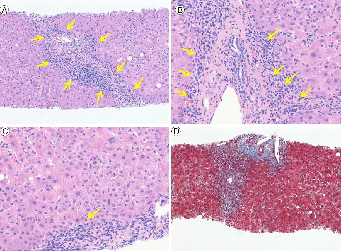
기저 질환으로 기관지확장증을 가진 58세 여자가 3주간 발열을 동반한 기침과 객혈 증상으로 호흡기내과에 입원하였다. 혈청 폐렴 마이코플라즈마 항체 양성으로 확인되어 마이코플라즈마 폐렴 진단으로 항생제(록시스로마이신 300 mg/day) 치료를 시작하였고, 이후 기침과 객혈 등의 증상은 호전되었다. 하지만 입원 3일째 혈액 검사에서 황달을 동반하지 않는 지속적인 혈청 아미노전이효소 증가(AST 418 IU/L, ALT 164 IU/L)를 보였고, 7일간 항생제를 복용 후 중단하였음에도 지속적인 혈청 아미노전이효소의 상승(AST 919 IU/L, ALT 987 IU/L)이 확인되었다. 복부 전산화단층촬영술에서는 간비대 이외의 특이 소견은 관찰되지 않았다. 환자는 정상 체중으로 음주력은 없었고 평소 복용 중인 약제도 없었다. 간염의 원인을 확인하기 위해 추가적인 혈액 검사를 시행하여 바이러스간염 등은 배제할 수 있었고, 항핵항체(antinuclear antibody, ANA) 1:80, 면역글로불린 G (immunoglobulin G, IgG) 2,376 mg/dL로 증가되어 자가면역간염의 진단을 위해 간 생검을 시행하였다. 조직 검사에서 중등도의 계면간염(interface hepatitis)과 형질세포 침투를 확인하였고(Fig. 1), 이를 반영한 개정된 International Autoimmune Hepatitis Group (IAIHG) 에 따른 자가면역간염 진단을 위한 점수는 16으로 명확한 자가면역간염에 합당하였다[4]. 치료로 경구 프레드니솔론 60 mg을 복용한 이후 혈청 아미노전이효소는 급격히 감소하였으며, 아자티오프린 추가 및 프레드니솔론 감량에도 혈청 아미노전이효소 및 IgG는 정상 범위까지 회복되었다.

자가면역간염의 병태생리 및 임상 소견

자가면역간염은 전 연령층과 모든 인종에서 발생할 수 있으며, 어른과 소아 모두 여성에서 호발(남여 비 약 1:3)하는 것으로 알려져 있다[5]. 추정 발병률은 지역과 발병 연령에 따라 다양하며, 유병률은 유럽 인구 100,000명당 11-25명이며 여성과 남성 모두에서 증가하고 있다[6]. 최근 연구에 따르면, 한국에서 자가면역간염은 중년(중위 연령 56세) 여성 (남녀 비 1:6.4)에서 호발하며, 유병률은 인구 100,000명당 4.82명이고 발생률은 인구 100,000명당 1.07로 확인되었다. 유럽이나 북미와 비교하여 적은 수로, 이는 환경적 및 유전적 차이로 인한 것으로 보인다[7].
자가면역간염은 유전적, 후성적, 면역학적, 환경적 요인의 상호 작용이 필요한 복합 유전 질환이다. 환경적 요인은 유전적 요인보다 면역 레퍼토리를 형성하는 데 중요한 역할을할 수 있으며, 특정한 환경적 요인(바이러스 감염이나 생체 이물에 대한 노출)은 유전적으로 자가면역간염에 취약한 사람에게서 자가 항원에 대한 자기 내성 상실을 유발할 수 있다[8].
자가면역간염은 무증상에서 급성/중증 또는 전격성 간염까지 임상적으로 다양하게 발현될 수 있다. 대부분의 자가면 역간염 환자는 피로, 권태감, 관절통 또는 무월경 등의 만성적인 비특이적 증상을 호소하며, 85%의 환자가 피로감을 주로 호소한다[9]. 진행성 만성 간질환의 징후(거미 모반, 비장 비대, 복수 등) 또는 간 외 자가면역 질환(백반증, 염증성 장질환)의 징후를 제외하고 신체적인 징후는 일반적으로 나타나지 않는다[10]. 또한 자가면역간염 환자의 25-34%는 무증상 환자이나 증상이 있는 환자와 유사한 조직학적 소견을 보이며, 2-120개월 이내에 증상이 발생하는 경우가 많으므로 증상의 부재를 이유로 치료를 시작하지 않아서는 안 된다[11-13].

자가면역간염의 진단 기준

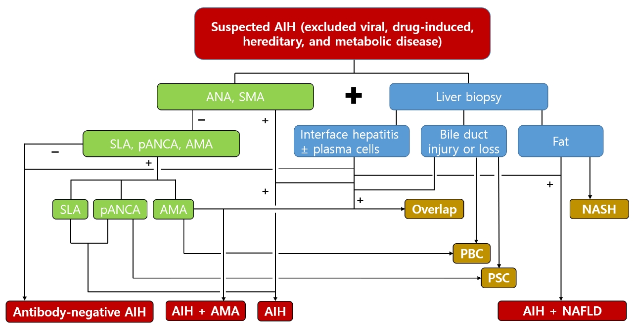
자가면역간염의 진단은 조직학적 이상(계면간염), 특징적인 임상 및 실험실 소견(증가된 혈청 AST, ALT 및 IgG) 그리고 하나 이상의 특징적인 자가항체의 존재를 기반으로 한다. 자가 면역간염은 특징적인 진단 표지자가 없고, 다른 만성 간질환의 원인(바이러스간염, 약제 유발 간 손상, 윌슨병, 유전성 혈색소 침착증 등)에 대한 배제가 필요하다(Fig. 2) [4,14,15]. 자가면역 간염에 대한 진단 기준과 진단 점수 체계는 1993년 제안되어 최근까지 개정되었고, 2015 유럽간학회 진료지침과 2019 미국간학회 진료지침에서도 언급되고 있다[3,15].

검사 결과

자가면역간염은 혈청 빌리루빈 농도와 아미노전이효소가 정상 범위보다 매우 증가되더라도 담즙정체효소는 정상 또는 미세한 상승 정도만 보이는 것이 특징적인 검사실 소견이다[4,16-18]. 다만 최근 연구에서 아미노전이효소의 상승과 함께 알칼리성 인산분해효소(alkaline phosphatase)가 아닌 감마 글루타밀전이효소(gamma glutamyl transferase)가 증가할 수도 있으며, 나아가 치료 반응의 독립적인 예측인자로 사용될 수 있음을 보고하였다[19,20].
간경변증이 동반되지 않은 자가면역간염 환자의 85%에서 혈청 IgG 증가가 확인되었으나 급성 발병 환자에서는 IgG 수치가 정상인 환자의 비율이 많았다[21,22]. 혈청 IgG가 높은 것은 자가면역간염의 특이적인 소견이며 혈청 IgA 및 IgM은 일반적으로 정상이다. 혈청 IgA와 IgM의 증가는 각각 알코올 지방간염 및 원발담즙성담관염과 같은 다른 질병이 있음을 시사한다[18].
아미노전이효소의 상승 정도는 조직학적으로 평가되는 자가면역간염의 중증도를 반영하지는 않지만, 아미노전이효소 및 면역글로불린 수치는 치료에 대한 반응과 관해 달성을 모니터링하는 데 유용한 지표로 이용된다[14].
바이러스 표지자의 부제가 자가면역간염을 진단하는 기준에 포함되지만, 바이러스간염 유병률이 높은 국가에서는 자가면역간염과 바이러스간염이 동반될 수 있다[23,24]. 이러한 경우, 명확한 진단 기준과 치료법이 정해진 바이러스간염과 비교하여 동반된 자가면역간염의 진단이 간과될 수 있기 때문에 주의가 필요하다. 자가면역간염을 치료하지 않으면 임상 증상의 악화 및 불량한 예후를 보일 수 있으므로, 임상적으로 자가면역간염이 의심될 때에는 적극적으로 혈청 학적 검사와 간 생검을 통해 동반된 자가면역간염이 있는지에 대한 평가를 고려하여야 한다.

자가항체

자가면역간염은 특정 자가항체에 따라 제1형과 제2형의두 가지 유형으로 나눌 수 있다(Table 1). 제1형은 자가면역 간염의 대부분을 차지하며 어느 연령에서나 발현하고 발견 당시 약 28% 정도에서 간경변증을 동반한다. 또한 치료에 대한 반응이 좋으나 약제 중단 시 재발의 가능성이 있다. 제2형은 주로 14세 미만의 소아에서 많이 발생하고, 급성의 임상 증상을 보이며 치료에 대한 반응이 좋지 않다. 자가항체의 경우, 제1형은 ANA 및/또는 평활근항체(smooth muscle antibody, SMA)/항액틴 항체(anti-actin antibody)를 특징으로 하고, 제2형은 대개 ANA 및 SMA의 부재와 항간신마이크로좀 1형 항체(antibody to liver kidney microsome type 1, anti-LKM1)를 특징으로 한다[25]. 이와 같이 ANA, SMA, anti-LKM-1 및 여러 자가항체는 자가면역간염 진단을 위한 중요한 지표이다[11]. ANA는 자가면역간염이 있는 북미 백인 성인의 80%에서 검출되며, SMA는 60%, anti-LKM-1은 3%에서 존재한다. 자가면역간염 환자의 49%는 진단 시 ANA, SMA 또는 anti-LKM-1를 단독 혈청학적 소견으로 보이며, 51%는 다중 항체에 대해 양성으로 확인된다[26]. 또한 특징적인 임상 소견이 동반된 자가면역간염의 최대 20%에서 ANA, SMA, anti-LKM-1 음성인 경우가 존재하므로, 혈청 음성 자가면역간염이 의심된다면 다른 자가 항체를 확인해 보아야 한다(Fig. 2). 알려진 다른 자가면역항체로는 perinuclear anti-neutrophil cytoplasmic antibodies (pANCA), liver specific cytosol antigen type 1 (anti-LC1), antibodies to asialoglycoprotein receptor (anti-ASGPR), IgA endomysial antibodies, antibodies to soluble liver antigen/liver pancreas (anti-SLA/LP), anti-actin 등이 있다(Table 2) [10,11,15]. 자가면역항체는 질병 특이적이 아니며 질병의 경과 중 변화할 수 있으므로, 한 번의 검사에서 자가항체 역가가 낮다고 자가면역간염을 배제하지 못한다. 또한 동반된 임상 소견이 없음에도 불구하고 항체 역가가 높다는 것만으로 자가면역간염을 진단 내릴 수 없다. 성인과 소아 모두에서 자가항체 역가가 질환의 진단 및 질병의 중등도와 치료 반응을 대략적으로 반영하지만, 질병의 경과나 예후에 대한 확립된 표지자는 아니다[27-30].

조직학적 특징

자가면역간염은 간 생검과 합당한 조직학적 소견 없이는 진단될 수 없다. 계면간염은 자가면역간염의 특징적인 조직학적 소견이며 66%에서 형질세포 침윤, 47%에서 소엽간염 (lobular hepatitis)이 동반된다. 중심소엽괴사(centrilobular necrosis)는 간경변증 동반 여부와 상관없이 29%에서 발견된다[31,32]. 또한 emperipolesis (active penetration by one cell into and through a large cell)는 65%의 자가면역간염에서 확인되며, rosettes는 33%에서 나타난다. 개별 조직학적 소견 중자가면역간염에 특이적인 소견은 없으나, 소엽으로 확장되는 문맥 림프구성 또는 림프형질구성 세포와의 계면 간염 소견, emperipolesis 및 rosettes 소견은 자가면역간염의 전형으로 간주된다[33]. 진단 시 조직학적 소견은 다른 간질환에 대한 진단을 배제하고, 염증 활성도의 중증도를 등급화하고 섬유화 정도를 나타내기 위해 필수적이다[34-36].

진단 점수 기준

자가면역간염의 전형적인 사례 외에 비전형적인 사례의 진단을 돕고, 진단을 정량화하여 객관적인 비교가 가능하도록 1993년에 IAIHG에 의해 점수제 진단이 고안되었고, 1999년수정 후 2008년에 간소화되었다(Tables 3, 4) [17,18]. 수정된 진단 점수 체계는 복잡하거나 특이한 특징을 가진 환자에게 선호되고, 간소화된 진단 점수 체계는 전형적인 환자에게 가장 정확하다. 간소화된 진단 점수 체계로 산정된 점수가 낮을수록, 수정된 진단 점수 체계로 재평가를 고려해야 한다[37]. 이러한 진단 점수 체계는 다른 원인으로 인한 간질환과 자가면역간염을 구별하는 데 매우 유용하며, 1) 자가면역 질환과의 연관성을 고려하여 자가면역 질환의 동반 여부, 2) 특정 HLA 표현형(DR3 or DR4), 3) pANCA 등과 같은 드문 자가항체 존재 유무와 함께 4) 치료 반응 및 재발 여부도 진단 항목에 반영된다. 또한 약제 복용 및 바이러스간염, 담즙 정체 질환, 알코올 간질환 등 타 질환의 감별을 도모하기 위한 항목들이 포함되어 있다. 이상과 같은 13개의 항목에 대한 점수를 더하여 치료 전 점수가 10-15점이면 가능성(probable), 16점 이상이면 확정적(definite) 진단을 내리며, 치료 후에는 각각 12-17점, 18점 이상이 필요하다[4]. IAIHG 점수 체계는 원인이 명확한 간질환과 자가면역간염을 구별하는 데 매우 유용하지만 전향적 연구를 통한 검증 부족, 동반된 간질환 (원발경화담관염, 원발담즙성담관염, 비알코올 지방간질환, 간이식, 전격성 간부전)에서의 정확성 부족, 다른 혈청학적 표지자가 제외된 점, 간접 면역 형광법에 의한 자가항체 측정과 같은 제한점 역시 존재한다[38,39].

자가면역간염의 치료

자가면역간염이 진단되면 치료를 고려해야 한다. 만약 치료하지 않으면 5년 및 10년 생존율이 50%와 10%에 불가할 정도로 예후가 불량하며, 심한 자가면역간염의 경우 6개월내 40%가 사망하며, 생존자 중 최소 40%가 간경변증으로 진행할 수 있다[40]. 하지만 면역억제제 등 적절한 치료를 할 경우 10년 생존율은 80-93%로 향상된다[41]. 자가면역간염에서 1차 요법의 목적은 약물 유발 합병증을 최소화하면서 증상을 개선하며, 간 내 염증을 조절하고 생화학적 관해 달성 및 추가적인 간질환의 진행을 예방하고 간 섬유화를 호전시키는 것이다. 이상적인 생화학적 관해 달성은 혈청 아미노전 이효소 및 IgG의 정상화이며, 비활동성 질환을 제외한 모든 자가면역간염 환자는 치료 대상이 될 수 있다[42,43]. 표준치료로 프레드니솔론 단독 또는 아자치오프린 병합요법을 고려할 수 있다. 아자치오프린의 경우 thiopurine methyltransferase (TPMT) genotyping을 통해 thiopurine methyltransferase의 결핍이 없는 것이 확인되면 프레드니솔론 치료 시작 2주 뒤 추가할 수 있다. 중증 급성 질환의 경우에는 고용량 프레드니솔론 단독으로 치료를 시작하고, 치료 시작 2주 후에 혈청 빌리루빈이 6 mg/dL 이하로 떨어지면 TPMT genotyping을 확인 후 아자티오프린을 추가할 수 있다[44].

결론

자가면역간염은 최근 점차 늘어나는 추세로, 이에 대한 진단의 중요성이 강조되고 있다. 자가면역간염의 진단은 우선 다른 만성 간질환에 대한 원인 배제가 필요하며, 특징적인 진단 표지자가 없기 때문에 급성이나 만성 간질환 환자에서 감마글로불린이 증가되어 있는 경우 자가항체를 검사함으로써 의심해볼 수 있다. 최근 자가면역성간염 및 원발담즙성담 관염이 의심되지만 ANA, AMA, SMA가 모두 음성인 경우 IgG 자가항체 4종(SLA/LP, LC-1, LKM-1, AMA-M2)을 추가로 시행할 수 있게 급여 기준이 변경되었으므로, 이와 같은 검사를 이용하는 것도 진단에 도움이 될 수 있다. 임상적으로 자가면역간염이 의심될 경우 간 생검은 자가면역간염의 진단에 필수적인 요소가 되고, 특징적인 조직학적 소견이 있을 경우 확정적으로 진단할 수 있다. 본고에서 기술한 바와 같이 자가면역간염의 임상 소견을 잘 이해하고 정확한 진단을 위해 적극적인 검사를 한다면 적절한 치료를 통해 환자의 예후를 호전시키는 것을 기대할 수 있겠다.

REFERENCES

1. Czaja AJ. Autoimmune hepatitis. Evolving concepts and treatment strategies. Dig Dis Sci 1995;40:435–456.
pmid

2. Vento S, Cainelli F. Is there a role for viruses in triggering autoimmune hepatitis? Autoimmun Rev 2004;3:61–69.
crossref

3. European Association for the Study of the Liver. EASL clinical practice guidelines: autoimmune hepatitis. J Hepatol 2015;63:971–1004.
crossref pmid

4. Alvarez F, Berg PA, Bianchi FB, et al. International Autoimmune Hepatitis Group report: review of criteria for diagnosis of autoimmune hepatitis. J Hepatol 1999;31:929–938.
crossref pmid

5. Ngu JH, Bechly K, Chapman BA, et al. Population-based epidemiology study of autoimmune hepatitis: a disease of older women? J Gastroenterol Hepatol 2010;25:1681–1686.
crossref pmid

6. Grønbæk L, Vilstrup H, Jepsen P. Autoimmune hepatitis in Denmark: incidence, prevalence, prognosis, and causes of death. A nationwide registry-based cohort study. J Hepatol 2014;60:612–617.
crossref pmid

7. Jeong SH. Current epidemiology and clinical characteristics of autoimmune liver diseases in South Korea. Clin Mol Hepatol 2018;24:10–19.
crossref pmid pmc

8. Floreani A, Leung PS, Gershwin ME. Environmental basis of autoimmunity. Clin Rev Allergy Immunol 2016;50:287–300.
crossref pmid

9. Czaja AJ. Natural history, clinical features, and treatment of autoimmune hepatitis. Semin Liver Dis 1984;4:1–12.
crossref pmid

10. Muratori P, Lalanne C, Fabbri A, Cassani F, Lenzi M, Muratori L. Type 1 and type 2 autoimmune hepatitis in adults share the same clinical phenotype. Aliment Pharmacol Ther 2015;41:1281–1287.
crossref pmid

11. Czaja AJ. Diagnosis and management of autoimmune hepatitis: current status and future directions. Gut Liver 2016;10:177–203.
crossref pmid pmc

12. Feld JJ, Dinh H, Arenovich T, Marcus VA, Wanless IR, Heathcote EJ. Autoimmune hepatitis: effect of symptoms and cirrhosis on natural history and outcome. Hepatology 2005;42:53–62.
crossref pmid

13. Muratori P, Lalanne C, Barbato E, et al. Features and progression of asymptomatic autoimmune hepatitis in Italy. Clin Gastroenterol Hepatol 2016;14:139–146.
crossref pmid

14. Manns MP, Czaja AJ, Gorham JD, et al. Diagnosis and management of autoimmune hepatitis. Hepatology 2010;51:2193–2213.
crossref pmid

15. Mack CL, Adams D, Assis DN, et al. Diagnosis and management of autoimmune hepatitis in adults and children: 2019 practice guidance and guidelines From the American Association for the Study of Liver Diseases. Hepatology 2020;72:671–722.
crossref pmid

16. Zachou K, Muratori P, Koukoulis GK, et al. Review article: autoimmune hepatitis -- current management and challenges. Aliment Pharmacol Ther 2013;38:887–913.
crossref pmid

17. Johnson PJ, McFarlane IG. Meeting report: International Autoimmune Hepatitis Group. Hepatology 1993;18:998–1005.
crossref pmid

18. Hennes EM, Zeniya M, Czaja AJ, et al. Simplified criteria for the diagnosis of autoimmune hepatitis. Hepatology 2008;48:169–176.
crossref pmid

19. Muratori P, Granito A, Quarneti C, et al. Autoimmune hepatitis in Italy: the Bologna experience. J Hepatol 2009;5:1210–1218.
crossref

20. Zachou K, Gatselis N, Papadamou G, Rigopoulou EI, Dalekos GN. Mycophenolate for the treatment of autoimmune hepatitis: prospective assessment of its efficacy and safety for induction and maintenance of remission in a large cohort of treatment-naïve patients. J Hepatol 2011;55:636–646.
crossref pmid

21. Fujiwara K, Fukuda Y, Yokosuka O. Precise histological evaluation of liver biopsy specimen is indispensable for diagnosis and treatment of acute-onset autoimmune hepatitis. J Gastroenterol 2008;43:951–958.
crossref pmid

22. Yasui S, Fujiwara K, Yonemitsu Y, Oda S, Nakano M, Yokosuka O. Clinicopathological features of severe and fulminant forms of autoimmune hepatitis. J Gastroenterol 2011;46:378–390.
crossref pmid

23. Georgiadou SP, Zachou K, Liaskos C, Gabeta S, Rigopoulou EI, Dalekos GN. Occult hepatitis B virus infection in patients with autoimmune liver diseases. Liver Int 2009;29:434–442.
crossref pmid

24. Rigopoulou EI, Zachou K, Gatselis N, Koukoulis GK, Dalekos GN. Autoimmune hepatitis in patients with chronic HBV and HCV infections: patterns of clinical characteristics, disease progression and outcome. Ann Hepatol 2013;13:127–135.
crossref pmid

25. Czaja AJ, Manns MP. The validity and importance of subtypes in autoimmune hepatitis: a point of view. Am J Gastroenterol 1995;90:1206–1211.
pmid

26. Czaja AJ. Performance parameters of the conventional serological markers for autoimmune hepatitis. Dig Dis Sci 2011;56:545–554.
crossref pmid

27. Czaja AJ. Behavior and significance of autoantibodies in type 1 autoimmune hepatitis. J Hepatol 1999;30:394–401.
crossref pmid

28. Czaja AJ, Homburger HA. Autoantibodies in liver disease. Gastroenterology 2001;120:239–249.
crossref pmid

29. Ben-Ari Z, Czaja AJ. Autoimmune hepatitis and its variant syndromes. Gut 2001;49:589–594.
crossref pmid pmc

30. Gregorio GV, McFarlane B, Bracken P, Vergani D, Mieli-Vergani G. Organ and non-organ specific autoantibody titres and IgG levels as markers of disease activity: a longitudinal study in childhood autoimmune liver disease. Autoimmunity 2002;35:515–519.
crossref pmid

31. Czaja AJ, Carpenter HA. Sensitivity, specificity, and predictability of biopsy interpretations in chronic hepatitis. Gastroenterology 1993;105:1824–1832.
crossref pmid

32. Miyake Y, Iwasaki Y, Terada R, et al. Clinical features of Japanese type 1 autoimmune hepatitis patients with zone III necrosis. Hepatol Res 2007;37:801–805.
crossref pmid

33. Balitzer D, Shafizadeh N, Peters MG, Ferrell LD, Alshak N, Kakar S. Autoimmune hepatitis: review of histologic features included in the simplified criteria proposed by the International Autoimmune Hepatitis Group and proposal for new histologic criteria. Mod Pathol 2017;30:773–783.
crossref pmid

34. Park SZ, Nagorney DM, Czaja AJ. Hepatocellular carcinoma in autoimmune hepatitis. Dig Dis Sci 2000;45:1944–1948.
pmid

35. Wang KK, Czaja AJ. Hepatocellular carcinoma in corticosteroid-treated severe autoimmune chronic active hepatitis. Hepatology 1988;8:1679–1683.
crossref pmid

36. Yeoman AD, Al-Chalabi T, Karani JB, et al. Evaluation of risk factors in the development of hepatocellular carcinoma in autoimmune hepatitis: implications for follow-up and screening. Hepatology 2008;48:863–870.
crossref pmid

37. Czaja AJ. Performance parameters of the diagnostic scoring systems for autoimmune hepatitis. Hepatology 2008;48:1540–1548.
crossref pmid

38. Yatsuji S, Hashimoto E, Kaneda H, Taniai M, Tokushige K, Shiratori K. Diagnosing autoimmune hepatitis in nonalcoholic fatty liver disease: is the International Autoimmune Hepatitis Group scoring system useful? J Gastroenterol 2005;40:1130–1138.
crossref pmid

39. Boberg KM, Chapman RW, Hirschfield GM, et al. Overlap syndromes: the International Autoimmune Hepatitis Group (IAIHG) position statement on a controversial issue. J Hepatol 2011;54:374–385.
crossref pmid

40. Soloway RD, Summerskill WH, Baggenstoss AH, et al. Clinical, biochemical, and histological remission of severe chronic active liver disease: a controlled study of treatments and early prognosis. Gastroenterology 1972;63:820–833.
crossref pmid

41. Cook GC, Mulligan R, Sherlock S. Controlled prospective trial of corticosteroid therapy in active chronic hepatitis. Q J Med 1971;40:159–185.
crossref pmid

42. Montano Loza AJ, Czaja AJ. Current therapy for autoimmune hepatitis. Nat Clin Pract Gastroenterol Hepatol 2007;4:202–214.
crossref pmid

43. Montano-Loza AJ, Carpenter HA, Czaja AJ. Improving the end point of corticosteroid therapy in type 1 autoimmune hepatitis to reduce the frequency of relapse. Am J Gastroenterol 2007;102:1005–1012.
crossref pmid

44. Czaja AJ, Carpenter HA. Decreased fibrosis during corticosteroid therapy of autoimmune hepatitis. J Hepatol 2004;40:646–652.
crossref pmid

Liver histology showing typical features of autoimmune hepatitis. (A) Lymphoplasmacytic inflammatory infiltration of the portal tract and interface hepatitis (arrows; hematoxylin and eosin [H&E], ×100). (B) Predominant lymphocytic portal infiltrate with clusters of plasma cells at interface (arrows; H&E, ×200). (C) A hepatocyte undergoing emperipolesis (active penetration by one cell into and through a large cell) (arrows; H&E, ×200) (D) Absent evidence of fibrosis (Masson Trichrome staining, ×100).
/upload/thumbnails/kjm-97-1-33f1.jpg
Figure 1.
Proposed diagnostic algorithm for the evaluation of suspected autoimmune hepatitis. AIH, autoimmune hepatitis; ANA, antinuclear antibodies; SMA, smooth muscle antibodies; SLA, soluble liver antigen; pANCA, perinuclear anti-neutrophil cytoplasmic antibodies; AMA, anti-mitochondrial antibodies; PBC, primary biliary cholangitis; PSC, primary sclerosing cholangitis; NSAH, nonalcoholic steatohepatitis; NAFLD, nonalcoholic fatty liver disease.
/upload/thumbnails/kjm-97-1-33f2.jpg
Figure 2.
Table 1.
Characteristic feature of type 1 and type 2 AIH
Features Type 1 AIH Type 2 AIH
Frequency US adults, 96% US children, 9-12%
UK children, 38%
Age at presentation Peripubertal and adults Usually under 14 years
Mode of presentation Chronic symptoms common Acute onset (~40%)
Ascites or GI bleeding rare Acute liver failure possible
Asymptomatic in 25-34% Relapse frequent
Acute in 25-75%
Acute severe in 2-6%
Laboratory features Hypergammaglobulinemia IgA levels may be reduced
Autoantibodies ANA, SMA, Antiactin, SLA Anti-LKM1 (anti-LC1, anti-LKM3)
Concurrent immune diseases Autoimmune thyroiditis Autoimmune thyroiditis
Rheumatic diseases Diabetes mellitus
IBD Vitilligo
Autoimmune overlap with PSC (ASC in children) Common in children Rare
Atypical pANCA-positive Atypical pANCA-negative
Overlap with PBC Seen in adults (not children) Not reported
Cirrhosis at presentation Adults, 28-33% (especially elderly) Rare
Children, ≤ 33%
Remission after drug withdrawal Possible Rare, usually need long-term immunosuppression

AIH, autoimmune hepatitis; GI, gastrointestinal; IgA, serum immunoglobulin A; ANA, antinuclear antibodies; SMA, smooth muscle antibodies; SLA, soluble liver antigen; LKM-1, liver kidney microsome type 1; LKM-3, liver kidney microsome type 3; LC1, liver specific cytosol antigen type 1; PSC, primary sclerosing cholangitis; ASC, autoimmune sclerosing cholangitis; pANCA, perinuclear anti-neutrophil cytoplasmic antibodies; PBC, primary biliary cholangitis.

Table 2.
Autoantibodies in the diagnosis of AIH
Antibody Target antigen Diagnostic value
ANA Chromatin, ribonucleoproteins Type 1 AIH
SMA Filamentous actin (F-actin), vimentin, desmin Type 1 AIH
LKM1 Cytochrome P450 2D6 (CYP2D6) Type 2 AIH
SLA Sep (O-phosphoserine) transfer RNA:Sec (selenocysteine) transfer RNA synthase Type 1 AIH
Severe AIH
Predicts relapse after treatment
Associated with poor outcome
p-ANCA (atypical) Β-tubulin isotype 5 Type 1 AIH
Nuclear lamina proteins PSC
ASC
Actin Filamentous (F) actin Type 1 AIH
α-Actinin Filamentous actin cross-linking proteins Investigational
Type 1 AIH
Prognostic biomarker
LKM3 UDP glucuronosyltransferase family 1 Type 2 AIH
Hepatitis D
LC-1 Formiminotransferase cyclodeaminase Type 2 AIH
LM Cytochrome P450 1A2 Dihydralazine-induced hepatitis
APECED hepatitis
AMA E2-subunits of pyruvate dehydrogenase complex PBC
PBC-AIH overlap syndrome
Type 1 AIH

AIH, autoimmune hepatitis; ANA, antinuclear antibodies; SMA, smooth muscle antibodies; LKM-1, liver kidney microsome type 1; SLA, soluble liver antigen; pANCA, perinuclear anti-neutrophil cytoplasmic antibodies; LKM-3, liver kidney microsome type 3; UDP, uridine diphosphate; LC1, liver specific cytosol antigen type 1; APECED, autoimmune polyendocrinopathy-candidias-ectodermal dystrophy; AMA, anti-mitochondrial antibodies; PBC, primary biliary cholangitis.

Table 3.
Original revised diagnostic scoring systems
Feature Score Feature Score
Gender Drug history
 Female + 2  Positive - 4
 Male 0  Negative + 1
ALP:AST (or ALT) Average alcohol intake
 < 1.5 - 2  < 25 g/day + 2
 1.5-3.0 0  > 60 g/day - 2
 > 3.0 + 2 Liver histology
Serum globulins or IgG > ULN  Interface hepatitis + 3
 > 2.0 + 3  Lymphoplasmacytic infiltrate + 1
 1.5-2.0 + 2  Rosetting of hepatocytes + 1
 1.0-1.5 + 1  None of above - 5
 < 1.0 0  Biliary changes - 3
ANA, SMA or LKM-1 (titers)  Other changes - 3
 > 1:80 + 3  Concurrent autoimmune diseases + 2
 1:80 + 2 Additional findings
 1:40 + 1  Other defined autoantibodies + 2
 < 1:40 0  HLA DRB1*03 or DRB1*04 + 1
 AMA (+) - 4 Response to therapy
Hepatitis viral markers  Complete + 2
 Positive - 3  Relapse + 3
 Negative + 3

Diagnostic scores
Pre-treatment score Post-treatment score
 Definite AIH > 15  Definite AIH > 17
 Probable AIH 10-15  Probable AIH 12-17
 Not AIH < 10  Not AIH < 12

AIH, autoimmune hepatitis; ALP, alkaline phosphatase; ALT, alanine aminotransferase; AMA, antimitochondrial antibodies; ANA, antinuclear antibodies; AST, aspartate aminotransferase; IgG, immunoglobulin G; LKM-1, liver kidney microsome type 1; SLA, soluble liver antigen; ULN, upper limit of normal range.

Table 4.
Simplified diagnostic scoring systems
Feature Value Score
ANA or SMA ≥ 1:40 titer + 1
ANA or SMA ≥ 1:80 titer + 1
LKM-1 ≥ 1:40 titer + 2
SLA Positive + 2
IgG > ULN + 1
> 1.1 × ULN + 2
Liver histology Compatible + 1
Typical + 2
Negative viral markers Yes + 2

Diagnostic Scores
≥ 6 Probable AIH
≥ 7 Definite AIH

AIH, autoimmune hepatitis; ANA, antinuclear antibodies; SMA, Smooth muscle antibodies; IgG, immunoglobulin G; LKM-1, liver kidney microsome type 1; SLA, soluble liver antigen.

TOOLS
METRICS Graph View
  • 1 Crossref
  •  0 Scopus
  • 4,803 View
  • 270 Download

Editorial Office
101-2501, Lotte Castle President, 109 Mapo-daero, Mapo-gu, Seoul 04146, Korea
Tel: +82-2-2271-6791    Fax: +82-2-790-0993    E-mail: kaim@kams.or.kr                

Copyright © 2024 by The Korean Association of Internal Medicine.

Developed in M2PI

Close layer
prev next